©2013 Baishideng Publishing Group Co.
World J Gastroenterol. Mar 21, 2013; 19(11): 1760-1769
Published online Mar 21, 2013. doi: 10.3748/wjg.v19.i11.1760
Published online Mar 21, 2013. doi: 10.3748/wjg.v19.i11.1760
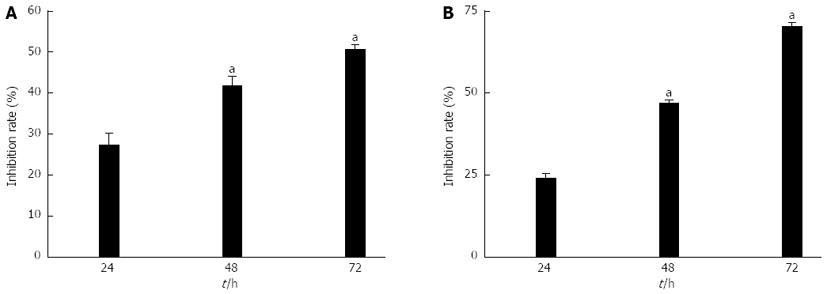
Figure 1 Reduced viability of SGC7901 and MGC803 cells after adenovirus Class I phosphoinositide 3-kinase-RNA interference-green fluorescent protein treatment.
A: SGC7901 cells (7 × 10⁴ cells/mL); B: MGC803 cells (7 × 10⁴ cells/mL) cultured with adenovirus Class I phosphoinositide 3-kinase [PI3K(I)]-RNA interference-green fluorescent protein (RNAi-GFP) (50 MOI) and adenovirus negative control-RNAi-GFP for 24, 48, and 72 h. Cell viability was analyzed by MTT assay. Values were given as mean ± SD of three independent experiments. aP < 0.05 vs control group.
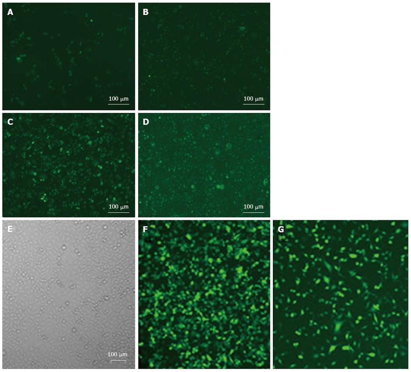
Figure 2 Transfection efficiency and cell morphology were detected by fluorescence microscope after adenovirus Class I phosphoinositide 3-kinase-RNA interference-green fluorescent protein and adenovirus negative control-RNA interference-green fluorescent protein treatment.
A-D: SGC7901 cells; E-G:MGC803 cells incubated with adenovirus Class I phosphoinositide 3-kinase [PI3K(I)]-RNA interference-green fluorescent protein (RNAi-GFP) (50 MOI) for the indicated time. A and E: Control group; B and F: 24 h after adenovirus PI3K(I)-RNAi-GFP treatment; C and G: 48 h adenovirus PI3K(I)-RNAi-GFP (50 MOI) treatment; D: 72 h after adenovirus PI3K(I)-RNAi-GFP (50 MOI) treatment (× 200) (n = 3).
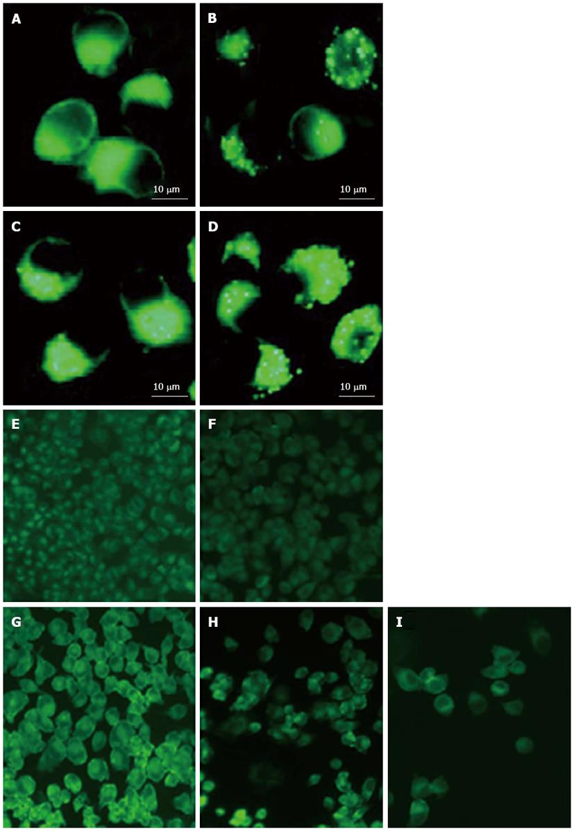
Figure 3 Monodansylcadaverine staining showed autophagy was activated after adenovirus Class I phosphoinositide 3-kinase-RNA interference-green fluorescent protein (50 MOI) treatment.
A-D: SGC7901 cells; E-I: MGC803 cells incubated with adenovirus Class I phosphoinositide 3-kinase [PI3K(I)]-RNA interference-green fluorescent protein (RNAi-GFP) (50 MOI) and adenovirus negative control (NC)-RNAi-GFP for the indicated time and stained with monodansylcadaverine (100 μmol/L). Fluorescence particles showed L-acoustic vector sensor. A and E: Control; B and F: Adenovirus NC-RNAi-GFP; C and G: 24 h after adenovirus PI3K(I)-RNAi-GFP (50 MOI) treatment (× 200) (n = 3); D and H: 48 h after adenovirus PI3K(I)-RNAi-GFP (50 MOI) treatment; I: 72 h after adenovirus PI3K(I)- RNAi-GFP (50 MOI) treatment (× 1000) (n = 3).
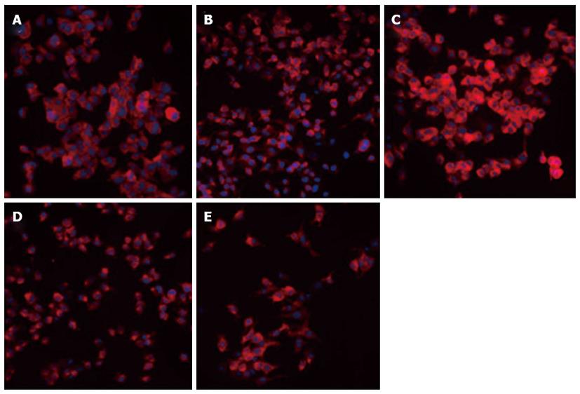
Figure 4 Microtubule-associated protein 1 light chain 3 expression and location in MGC803 cells after treatment with adenovirus Class I phosphoinositide 3-kinase-RNA interference-green fluorescent protein.
Cells were treated with adenovirus Class I phosphoinositide 3-kinase [PI3K(I)]-RNA interference-green fluorescent protein (RNAi-GFP) (50 MOI) for 24 h (C), 48 h (D), and 72 h (E), and analyzed with an immunofluorescence microscope. A: Control; B: Adenovirus negative control-RNAi-GFP (× 400) (n = 3). Adenovirus PI3K(I)-RNAi-GFP increased the punctate distribution of light chain 3 from 24 to 72 h.
Figure 5 Flow cytometric analysis of mitochondria membrane potential in the control and adenovirus Class I phosphoinositide 3-kinase-RNA interference-green fluorescent protein-treated SGC7901 cells.
A: Control: adenovirus negative control RNA interference-green fluorescent protein (RNAi-GFP), cells were treated with adenovirus Class I phosphoinositide 3-kinase [PI3K(I)]-RNAi-GFP (50 MOI) for 24 h (B), 48 h (C) and 72 h (D), and were then stained with JC-1 (5 μmol/L) for 30 min.
Figure 6 Effects of adenovirus Class I phosphoinositide 3-kinase-RNA interference-green fluorescent protein on light chain 3 and Beclin-1 protein expression/Bcl-2 and p53 protein expression in SGC7901 cells.
A: Effect of adenovirus Class I phosphoinositide 3-kinase [PI3K(I)]-RNA interference-green fluorescent protein (RNAi-GFP) (50 MOI) and adenovirus negative control (NC)-RNAi-GFP on light chain 3 (LC3) and Beclin-1 protein expression. Control: Adenovirus NC-RNAi-GFP. SGC7901 cells were treated with adenovirus PI3K(I)-RNAi-GFP (50 MOI) for 24 to 72 h then harvested for the extraction of total proteins. Adenovirus PI3K(I)-RNAi-GFP up-regulates the expression of LC3 and Beclin protein; B: Adenovirus PI3K(I)-RNAi-GFP (50 MOI) and adenovirus NC-RNAi-GFP on Bcl-2 and p53 protein expression. Control: Adenovirus NC-RNAi-GFP. SGC7901 cells were treated with adenovirus PI3K(I)-RNAi-GFP (50 MOI) for 24 to 72 h then harvested for the extraction of total proteins. Adenovirus PI3K(I)-RNAi-GFP up-regulates the expression of p53 and down-regulates the expression of Bcl-2 protein.
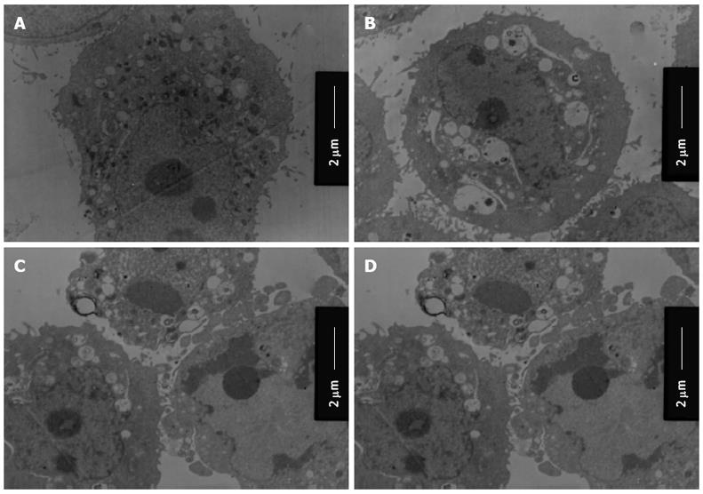
Figure 7 Ultrastructure of SGC7901 cells undergo autophagy, apoptosis and necrosis after adenovirus Class I phosphoinositide 3-kinase-RNA interference-green fluorescent protein treatment.
A: Control: Adenovirus negative control RNA interference-green fluorescent protein (RNAi-GFP); B: Adenovirus Class I phosphoinositide 3-kinase [PI3K(I)]-RNAi-GFP (50 MOI)-treated (24 h); C: Adenovirus PI3K(I)-RNAi-GFP (50 MOI)-treated (48 h); D: Adenovirus PI3K(I)-RNAi-GFP (50 MOI)-treated (72 h).
- Citation: Zhu BS, Yu LY, Zhao K, Wu YY, Cheng XL, Wu Y, Zhong FY, Gong W, Chen Q, Xing CG. Effects of small interfering RNA inhibit Class I phosphoinositide 3-kinase on human gastric cancer cells. World J Gastroenterol 2013; 19(11): 1760-1769
- URL: https://www.wjgnet.com/1007-9327/full/v19/i11/1760.htm
- DOI: https://dx.doi.org/10.3748/wjg.v19.i11.1760


