©2013 Baishideng Publishing Group Co.
World J Gastroenterol. Mar 21, 2013; 19(11): 1683-1698
Published online Mar 21, 2013. doi: 10.3748/wjg.v19.i11.1683
Published online Mar 21, 2013. doi: 10.3748/wjg.v19.i11.1683
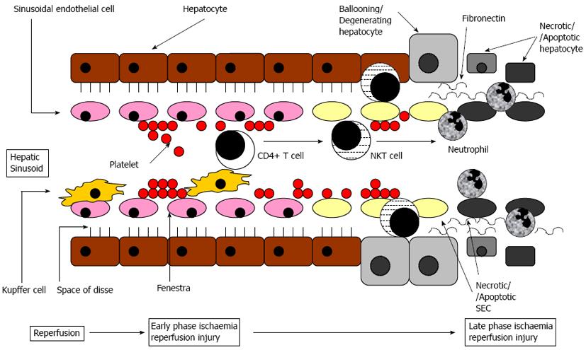
Figure 1 Schematic diagram of cellular mechanisms of liver ischaemia reperfusion injury within a liver sinusoid and the surrounding area containing hepatocytes.
Initial sinusoidal perfusion failure from platelet plugging, then Kupffer cells activate CD4+ T cells that activate natural killer T (NKT) cell which cause sinusoidal endothelial cells (SEC) and hepatocyte injury, followed by neutrophil activation, adhesion and transmigration causing more cell injury.
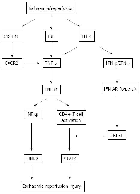
Figure 2 Cytokine and downstream signalling pathways in liver ischaemia reperfusion injury.
Following liver ischaemia reperfusion, there is activation of tumor necrosis factor-α (TNF-α) is by chemokine (CXCL) 10, interferon regulatory factor (IRF) and toll-like receptor (TLR) 4 in parallel. TNF-α activates downstream hepatocyte/sinusoidal endothelial cells (SEC) nuclear factor κβ (NFκβ) and CD4+ T cells separately which activate c-Jun N-terminal protein kinase-2 (JNK-2) and signal transducer activator of transcription-4 (STAT4), respectively leading to increased cell injury. A parallel pathway of cell injury occurs where TLR4 activation stimulates interferon (IFN)-β and IFN-γ expression, which acting through their receptor IFN receptor subtype (AR) activate interferon regulatory element (IRE)-1 which in turn activate CD4+ T cells. CXCR: Chemokine receptor.
- Citation: Datta G, Fuller BJ, Davidson BR. Molecular mechanisms of liver ischemia reperfusion injury: Insights from transgenic knockout models. World J Gastroenterol 2013; 19(11): 1683-1698
- URL: https://www.wjgnet.com/1007-9327/full/v19/i11/1683.htm
- DOI: https://dx.doi.org/10.3748/wjg.v19.i11.1683


