Copyright
©2013 Baishideng Publishing Group Co.
World J Gastroenterol. Mar 14, 2013; 19(10): 1665-1668
Published online Mar 14, 2013. doi: 10.3748/wjg.v19.i10.1665
Published online Mar 14, 2013. doi: 10.3748/wjg.v19.i10.1665
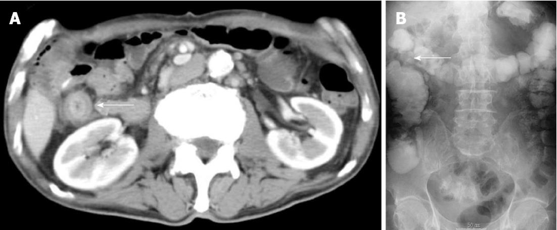
Figure 1 Examinations at seven years after pancreatoduodenectomy.
A: An abdominal computed tomography showing thickening of the ascending colon wall with enhancement and luminal narrowing (arrow); B: A contrast examination showing a 5-cm stricture of the ascending colon (arrow).
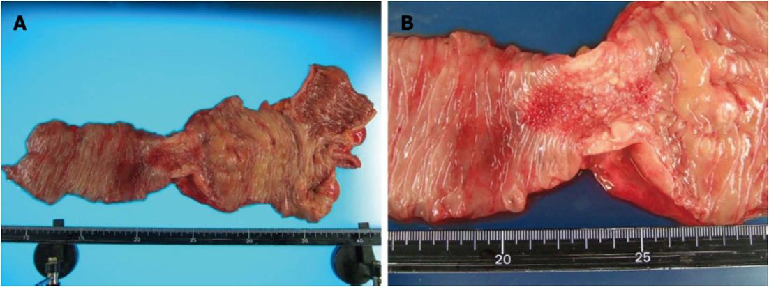
Figure 2 A pathological specimen showing a 5-cm wall thickening and a cobblestone-like appearance of the ascending colon.
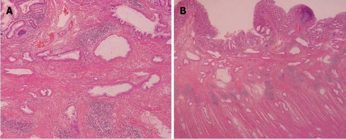
Figure 3 Histological findings revealed the cancer nests invading from the subserosa to the muscular and submucosal layers of the colon.
A: × 4; B: × 10.
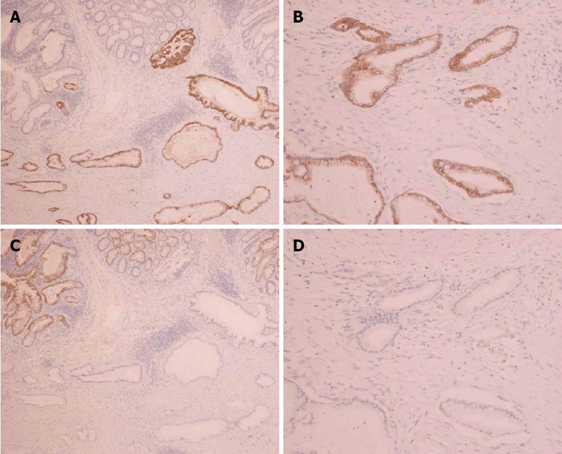
Figure 4 Photomicrographs (× 10).
A: Immunostaining showing cytokeratin 7 positivity in colonic metastasis; B: Immunostaining showing cytokeratin 20 negativity in colonic metastasis; C: Immunostaining showing cytokeratin 7 positivity in primary pancreatic cancer; D: Immunostaining showing cytokeratin 20 negativity in primary pancreatic cancer.
- Citation: Inada K, Shida D, Noda K, Inoue S, Warabi M, Umekita N. Metachronous colonic metastasis from pancreatic cancer seven years post-pancreatoduodenectomy. World J Gastroenterol 2013; 19(10): 1665-1668
- URL: https://www.wjgnet.com/1007-9327/full/v19/i10/1665.htm
- DOI: https://dx.doi.org/10.3748/wjg.v19.i10.1665


