Copyright
©2013 Baishideng Publishing Group Co.
World J Gastroenterol. Jan 7, 2013; 19(1): 133-136
Published online Jan 7, 2013. doi: 10.3748/wjg.v19.i1.133
Published online Jan 7, 2013. doi: 10.3748/wjg.v19.i1.133
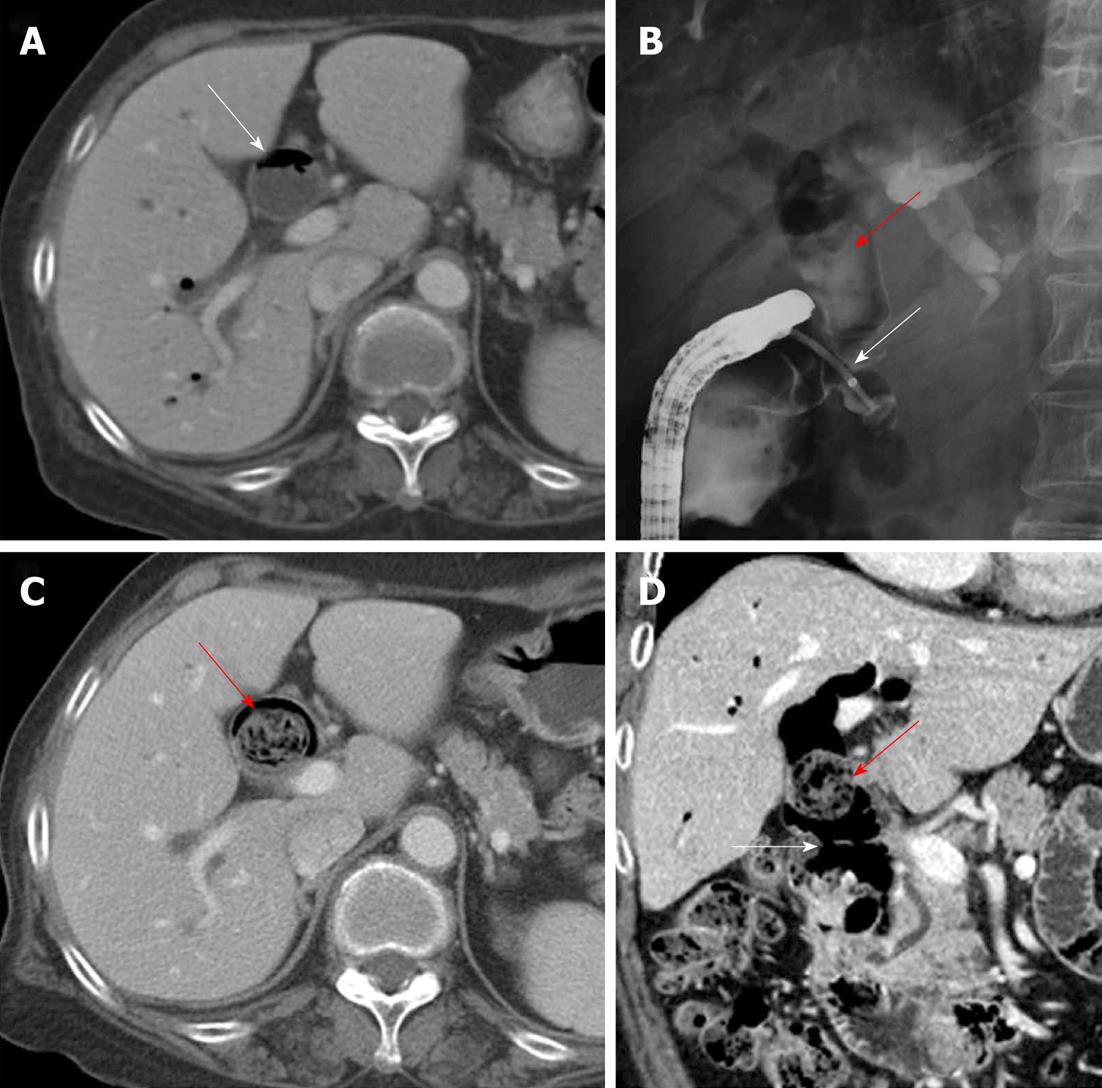
Figure 1 Development of a biliary phytobezoar in the extrahepatic bile duct.
A: Axial portal venous-phase computed tomography (PVP CT) 3 years ago showing diffuse bile duct dilatation (white arrow) with pneumobilia; B: On endoscopic retrograde cholangiopancreatography, there was no abnormal filling defect except pneumobilia (red arrow) in the dilated extrahepatic bile duct (EHD). A choledochoduodenal fistula (white arrow) in the distal common bile duct which was cannulated by endoscopy; C and D: Axial PVP CT and its coronal reformatted image at 6-mo follow-up showed a dilated EHD and a newly developed intraductal ovoid mass with interstitial air (red arrows), as well as the choledochoduodenalfistula (white arrow).
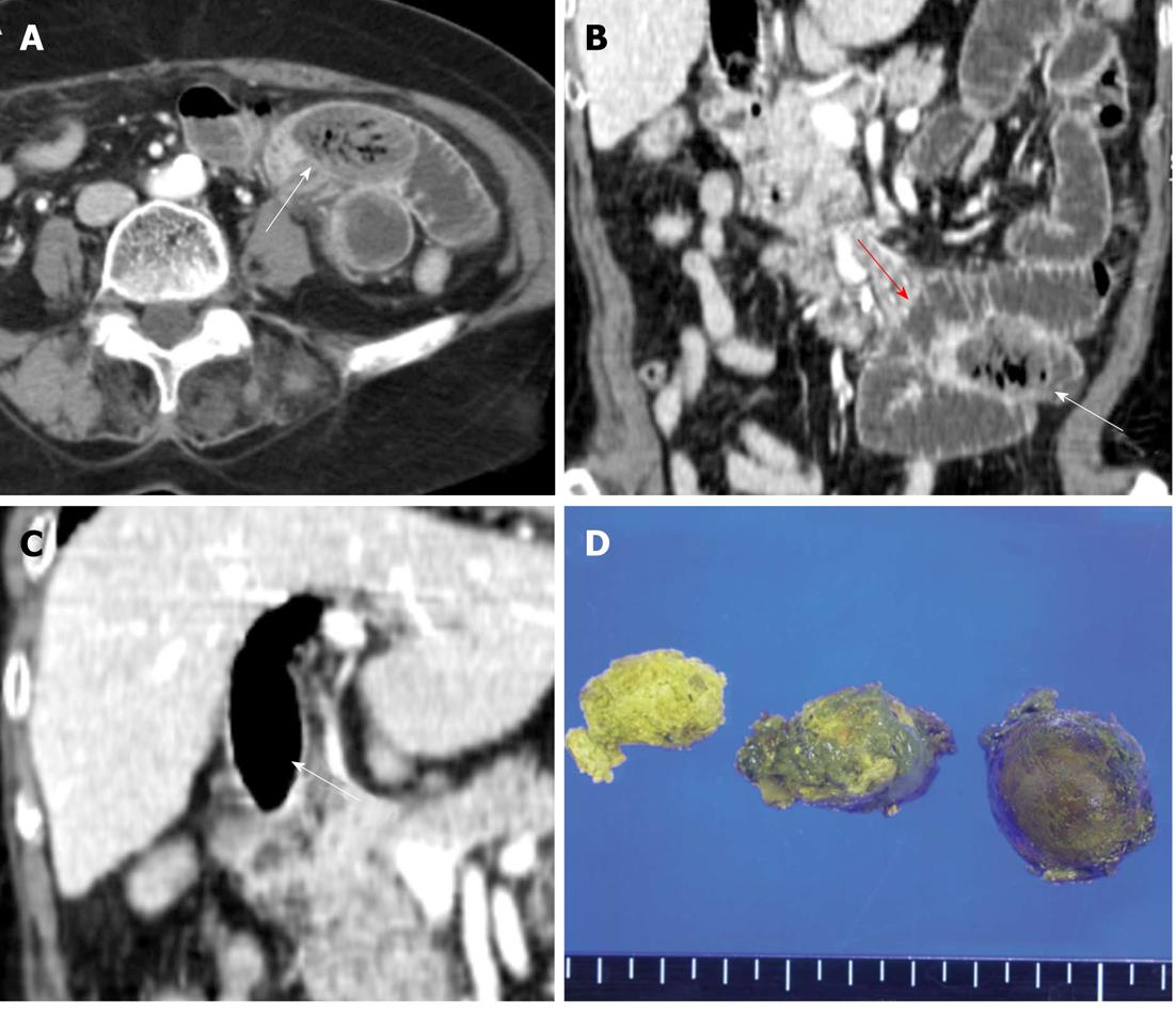
Figure 2 Migration of the biliary phytobezoar resulting in mechanical obstruction of the jejunum.
A and B: Axial portal venous computed tomography and coronal reformatted image at admission showed the migration of the ovoid intraluminal mass (white arrows) with mottled gas pattern to the jejunal loop, as well as a dilated proximal loop (red arrow) containing air-fluid levels suggestive of obstruction; C: Empty extrahepatic bile duct (white arrow) with extensive residual pneumobilia after migration of the bezoar; D: Gross specimen and cut surface of the biliary bezoar.
- Citation: Kim Y, Park BJ, Kim MJ, Sung DJ, Kim DS, Yu YD, Lee JH. Biliary phytobezoar resulting in intestinal obstruction. World J Gastroenterol 2013; 19(1): 133-136
- URL: https://www.wjgnet.com/1007-9327/full/v19/i1/133.htm
- DOI: https://dx.doi.org/10.3748/wjg.v19.i1.133


