©2012 Baishideng Publishing Group Co.
World J Gastroenterol. Mar 7, 2012; 18(9): 952-959
Published online Mar 7, 2012. doi: 10.3748/wjg.v18.i9.952
Published online Mar 7, 2012. doi: 10.3748/wjg.v18.i9.952
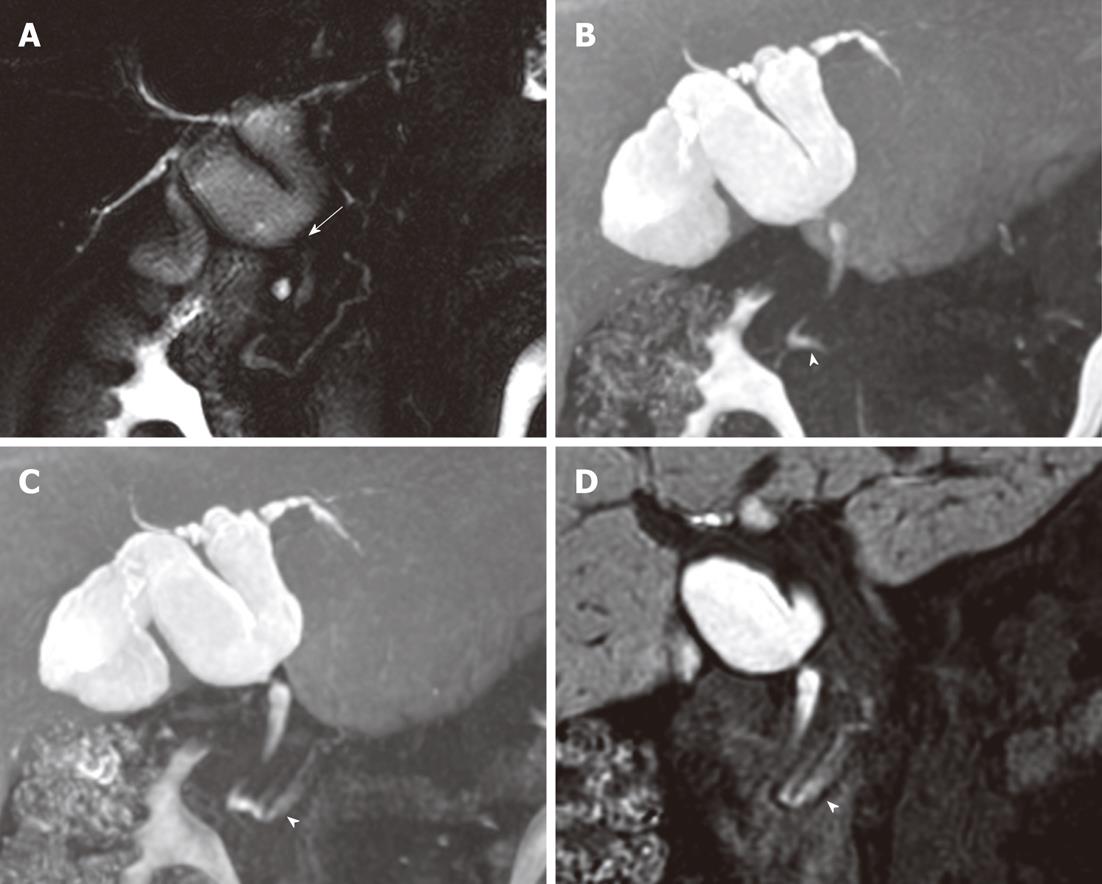
Figure 1 A 52-year-old woman with anomalous union of the pancreatico-biliary duct and a type Ia choledochal cyst.
A: Fusiform dilation of the common hepatic and cystic ducts with a focal stricture in the common bile duct (arrow) on T2-magnetic resonance cholangiography (MRC); B: Maximum intensity projection (MIP) reconstruction image of 60-min delayed gadoxetic acid-enhanced MRC shows the main pancreatic duct (arrowhead), indicating biliopancreatic reflux; C: MIP reconstruction image of 30-min delayed gadoxetic acid-enhanced MRC after a fatty meal shows progression of contrast media (arrowhead) along the main pancreatic duct; D: Gadoxetic acid-enhanced MRC coronal image taken 30 min after a fatty meal shows visualization of the main pancreatic duct (arrowhead) using contrast material.
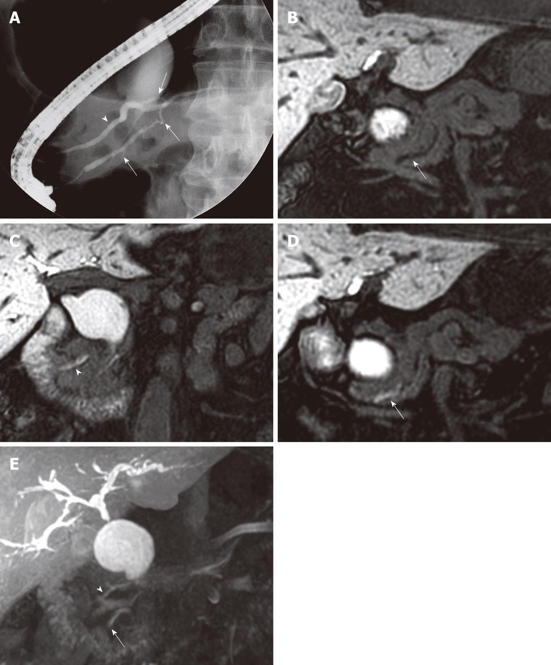
Figure 2 A 64-year-old man with anomalous union of the pancreatico-biliary duct and a type Ia choledochal cyst.
A: Endoscopic retrograde cholangiopancreatography shows cystic dilation of the extrahepatic bile duct with a focal stricture in the distal common bile duct. The end of the long ventral pancreatic duct (duct of Wirsung, arrows) is fused with the dorsal pancreatic duct (duct of Santorini, arrowhead). The common bile duct inserts into the ventral pancreatic duct; B: Sixty-minute delayed gadoxetic acid-enhanced magnetic resonance cholangiography (MRC) coronal image does not visualize the main pancreatic duct (arrow); C and D: Gadoxetic acid-enhanced MRC coronal images taken 30 min after a fatty meal show the duct of Santorini (arrowhead) and duct of Wirsung (arrow), indicating biliopancreatic reflux of contrast material; E: Gadoxetic acid-enhanced MRC maximum intensity projection reconstruction images taken 30 min after a fatty meal show major and minor pancreatic ducts.
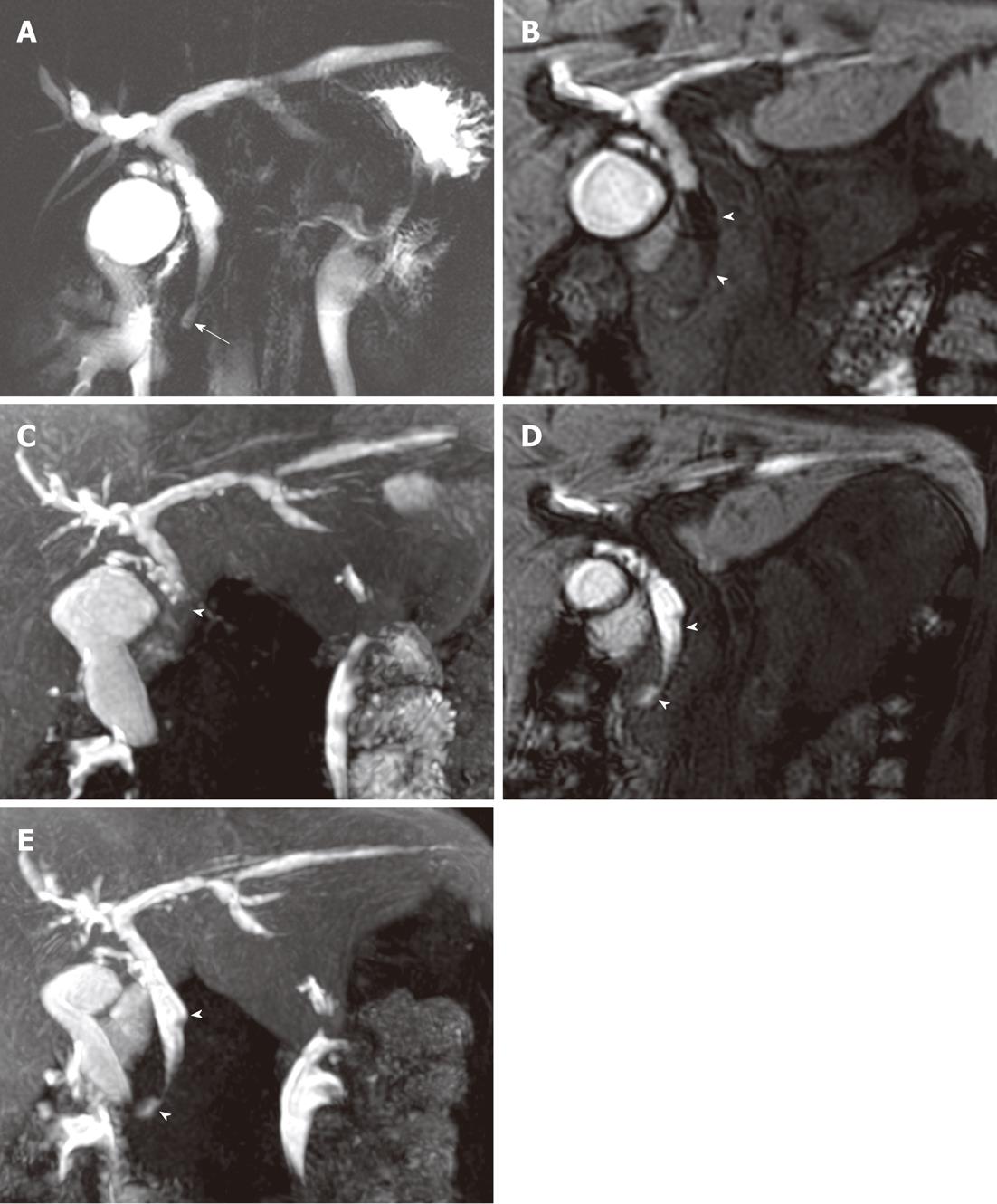
Figure 3 A 40-year-old woman with anomalous union of the pancreatico-biliary duct and a type Ic choledochal cyst.
A: T2-magnetic resonance cholangiography (MRC) shows a long common channel (arrow), with diffuse bile duct dilation; B: Sixty-minute delayed gadoxetic acid-enhanced MRC coronal image; C: Maximum intensity projection (MIP) reconstruction image show a filling defect (arrow heads) in the central portion of the distal common bile duct (CBD); D: Gadoxetic acid-enhanced MRC coronal images taken 30 min after a fatty meal; E: MIP reconstruction images show a decreased filling defect (arrow heads) in the distal CBD, indicative of pancreatico-biliary reflux.
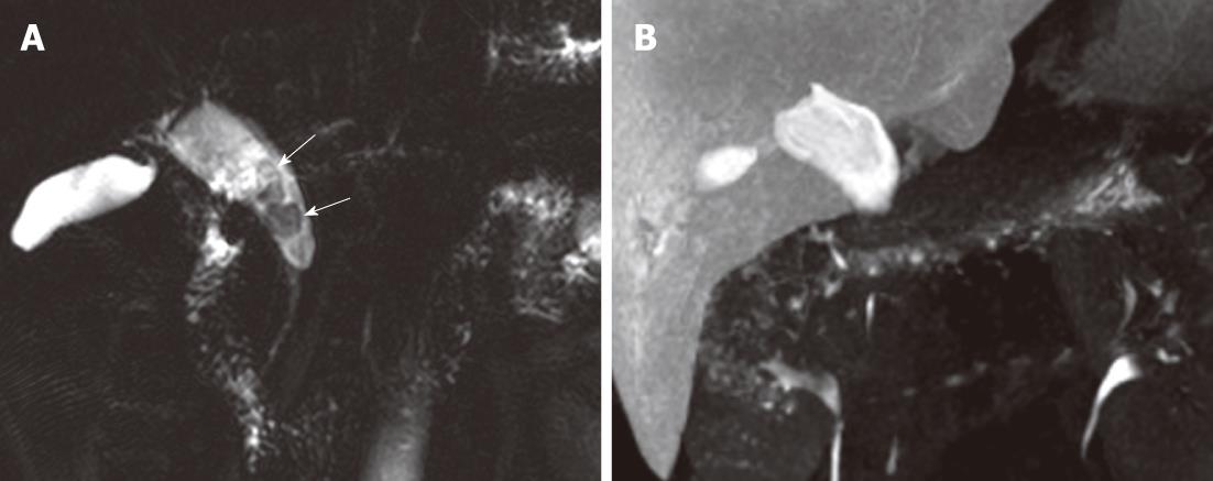
Figure 4 A 35-year-old woman with a type Ia choledochal cyst and multiple common bile duct stones.
A: Fusiform dilatation of the extrahepatic bile duct is seen on T2-magnetic resonance cholangiography (MRC) and multiple nodular filling defects (arrows) are seen in the common bile duct (CBD) which represent CBD stones; B: Contrast media did not pass through the ampulla of Vater until 30 min after fatty meal ingestion and multiple CBD stones are not seen due to masking by contrast media with high signal intensity.
- Citation: Yeom SK, Lee SW, Cha SH, Chung HH, Je BK, Kim BH, Hyun JJ. Biliary reflux detection in anomalous union of the pancreatico-biliary duct patients. World J Gastroenterol 2012; 18(9): 952-959
- URL: https://www.wjgnet.com/1007-9327/full/v18/i9/952.htm
- DOI: https://dx.doi.org/10.3748/wjg.v18.i9.952


