©2012 Baishideng Publishing Group Co.
World J Gastroenterol. Mar 7, 2012; 18(9): 905-914
Published online Mar 7, 2012. doi: 10.3748/wjg.v18.i9.905
Published online Mar 7, 2012. doi: 10.3748/wjg.v18.i9.905
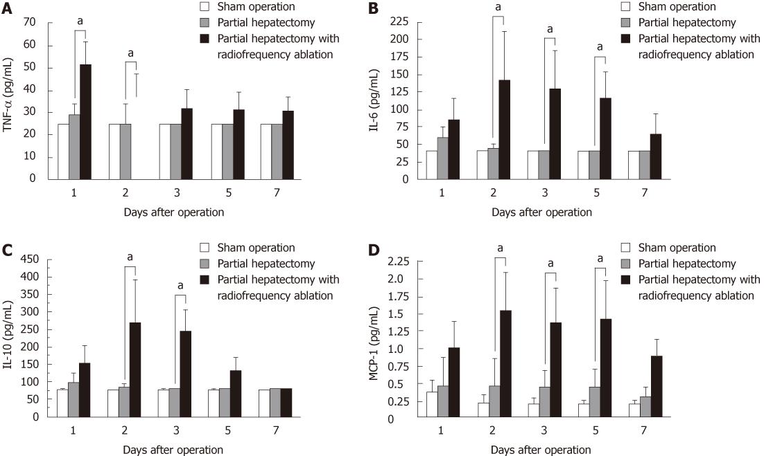
Figure 1 Changes in serum tumor necrosis factor-α (A), IL-6 (B), IL-10 (C), and monocyte chemoattractant protein-1 (D) levels after the operations.
aP < 0.05 between groups. TNF-α: Tumor necrosis factor-α; MCP-1: Monocyte chemoattractant protein-1.
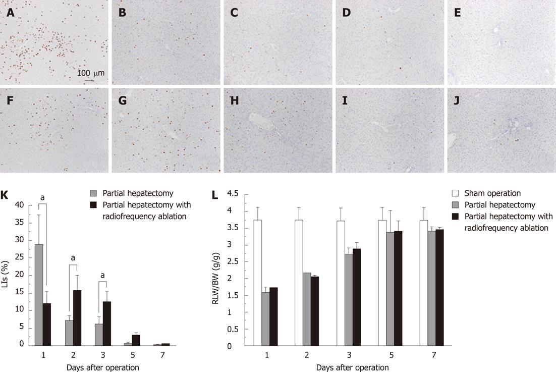
Figure 2 Immunohistochemistry for 5-bromo-2-deoxyuridine in the partial hepatectomy group (A-E) and in the partial hepatectomy with radiofrequency ablation group (F-J) at 1 d (A and F), 2 d (B and G), 3 d (C and H), 5 d (D and I) and 7 d (E and J) after the operations.
K: Comparison of labeling indices in the partial hepatectomy group and partial hepatectomy with radiofrequency ablation group; L: The ratio of liver weight/body weight shows restitution of the remnant liver. aP < 0.05 between groups. LIs: Labeling indices; RLW/BW: Residual liver weight/body weight.
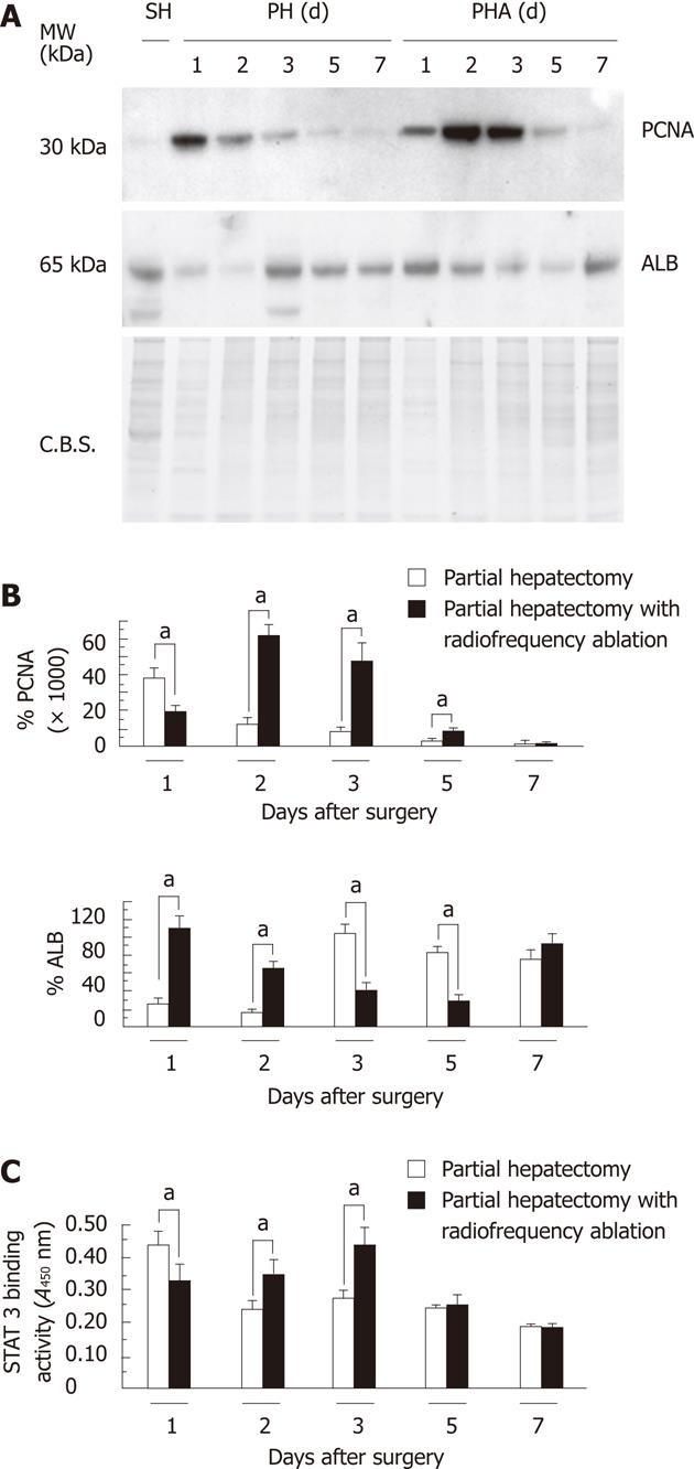
Figure 3 Proliferation cell nuclear antigen expression in the nuclear protein and albumin expression in the cytosol after the operations.
A: Western blotting analysis for proliferation cell nuclear antigen expression (PCNA); B: Densitometric analysis of each protein signal; C: STAT3 DNA-binding activity in the nuclear protein after the operations. aP < 0.05 between groups. SH: Sham hepatectomy; PH: Partial hepatectomy; PHA: Partial hepatectomy with radiofrequency ablation; MW: Molecular weight; C.B.S.: Coomassie blue staining; ALB: Albumin.
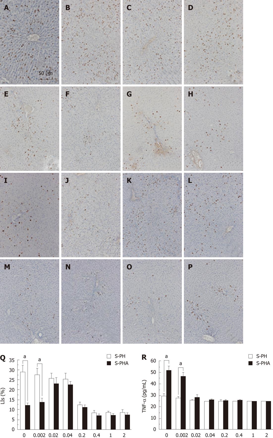
Figure 4 Immunohistochemistry for 5-bromo-2-deoxyuridine staining in the partial hepatectomy with dexamethasone pretreatment group (A-H) and partial hepatectomy after radiofrequency ablation with dexamethasone pretreatment group (I-P) at 1 d after the operations.
Animals were pretreated with 0 mg/kg (A and I), 0.002 mg/kg (B and J), 0.02 mg/kg (C and K), 0.04 mg/kg (D and L), 0.2 mg/kg (E and M), 0.4 mg/kg (F and N), 1 mg/kg (G and O), or 2 mg/kg (H and P) dexamethasone. Q: The brown nuclei are positive for 5-bromo-2-deoxyuridine. The labeling indices were calculated from 20 fields in three different sections per treatment for five different animals; R: Serum tumor necrosis factor-α levels at 1 d after the operations. The horizontal axis presents each concentration of dexamethasone pretreatment. aP < 0.05 between groups. S-PH: Steroid pretreatment in the partial hepatectomy group; S-PHA: Steroid pretreatment in the partial hepatectomy after radiofrequency ablation group; LIs: Labeling indices.
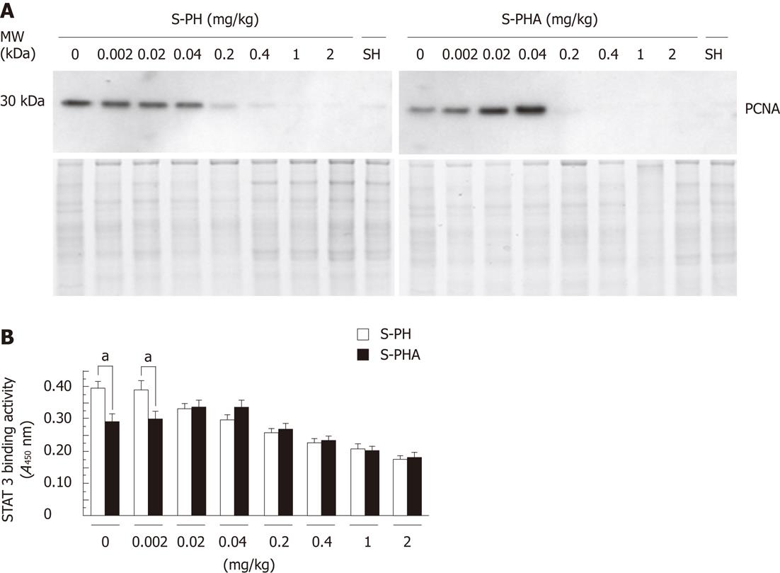
Figure 5 Proliferation cell nuclear antigen expression in the nuclear protein at 1 d after the operations.
A: Western blotting analysis for proliferation cell nuclear antigen (PCNA) expression. Animals were pretreated with various concentrations of dexamethasone 30 min prior to the hepatectomy; B: STAT3 DNA-binding activity in the nucleus at 1 d after the operations. aP < 0.05 between groups. S-PH: Steroid pretreatment in the partial hepatectomy group; S-PHA: Steroid pretreatment in the partial hepatectomy after radiofrequency ablation group; MW: Molecular weight.
- Citation: Shibata T, Mizuguchi T, Nakamura Y, Kawamoto M, Meguro M, Ota S, Hirata K, Ooe H, Mitaka T. Low-dose steroid pretreatment ameliorates the transient impairment of liver regeneration. World J Gastroenterol 2012; 18(9): 905-914
- URL: https://www.wjgnet.com/1007-9327/full/v18/i9/905.htm
- DOI: https://dx.doi.org/10.3748/wjg.v18.i9.905


