©2012 Baishideng Publishing Group Co.
World J Gastroenterol. Mar 7, 2012; 18(9): 889-895
Published online Mar 7, 2012. doi: 10.3748/wjg.v18.i9.889
Published online Mar 7, 2012. doi: 10.3748/wjg.v18.i9.889
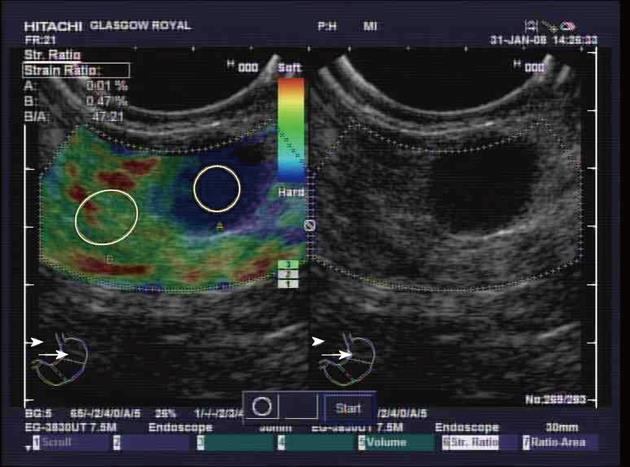
Figure 1 Endoscopic ultrasound image of a malignant appearing lymph node.
The right-hand side of the image displays all 4 of the conventional endoscopic ultrasound criteria characteristics of malignant nodes with regard to size (> 1 cm), shape (round), density (hypodense) and distinction of border (clear edge). The left-hand side of the image is a superimposed elastographic image with strain ratio measurement between an area of the lymph node and a surrounding area of tissue.
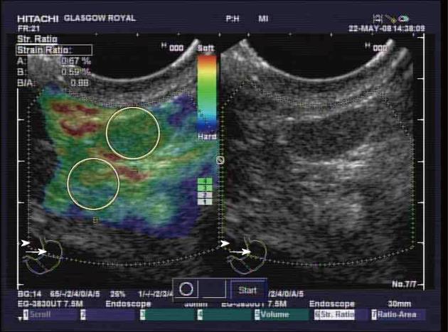
Figure 2 Endoscopic ultrasound elastography of a benign lymph node.
The right-hand side of the image displays standard grey-scale endoscopic ultrasound images while on the left is a superimposed elastography image. In the elastography image window the strain ratio measurements of the two areas outlined in the yellow circles is shown as a percentage in the top left-hand corner. The calculated strain ratio is shown as B/A. The elastographic signal is indicated by the bar column in the bottom right of the elastographic image window.
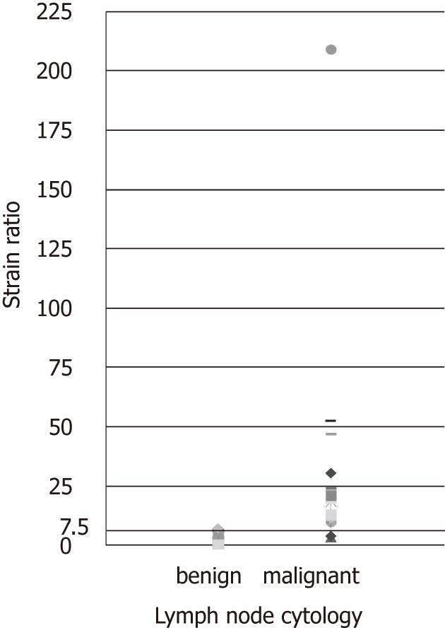
Figure 3 Plot of elastography strain ratio for cytologically proven benign or positive lymph nodes.
The cut-off line of ≥ 7.5 is the optimal strain ratio for discriminating between benign and malignant lymph nodes.
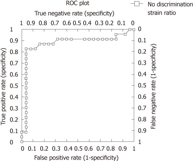
Figure 4 Receiver operating characteristic curve for elastography strain ratio.
The receiver operating characteristic area under the curve was 0.87 (P < 0.0001).
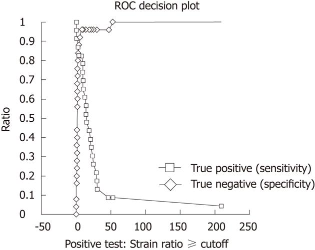
Figure 5 Receiver operating characteristic sensitivity, specificity based decision plot to determine the optimal elastography strain ratio cut-off point.
The sensitivity and specificity lines cross at strain ratio 7.5.
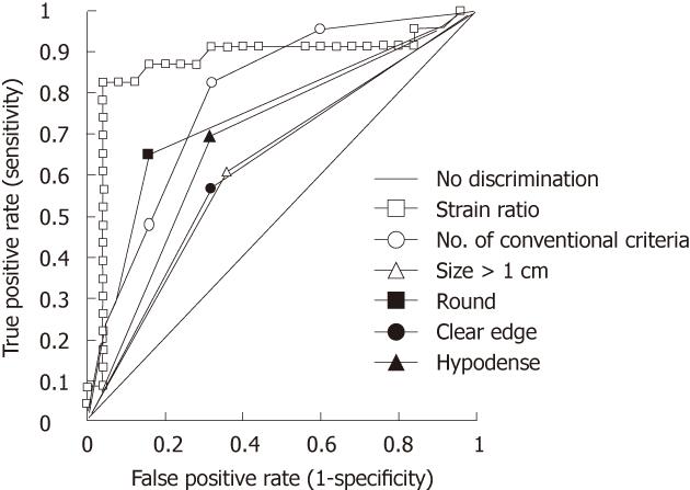
Figure 6 Receiver operating characteristic curve comparison of elastography strain ratio against conventional endoscopic ultrasound criteria both in combination and individually (size > 1 cm, round, hypodense, clear edge).
- Citation: Paterson S, Duthie F, Stanley AJ. Endoscopic ultrasound-guided elastography in the nodal staging of oesophageal cancer. World J Gastroenterol 2012; 18(9): 889-895
- URL: https://www.wjgnet.com/1007-9327/full/v18/i9/889.htm
- DOI: https://dx.doi.org/10.3748/wjg.v18.i9.889


