Copyright
©2012 Baishideng Publishing Group Co.
World J Gastroenterol. Nov 28, 2012; 18(44): 6427-6436
Published online Nov 28, 2012. doi: 10.3748/wjg.v18.i44.6427
Published online Nov 28, 2012. doi: 10.3748/wjg.v18.i44.6427
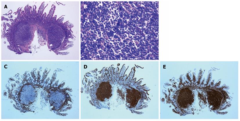
Figure 1 Typical histological features of follicular lymphoma.
A, B: Small cleaved cells that infiltrated the duodenal mucosa and formed lymphoid follicles are present (hematoxylin and eosin staining); C: Representative immunohistochemical staining of lymphoma cells negative for CD3; D: Lymphoma cells positive for CD10; E: Lymphoma cells positive for BCL-2. All of the images shown are at 40 × magnification, except for panel B which is at 400 × magnification.
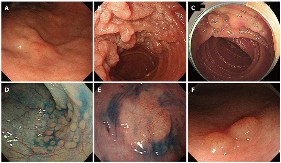
Figure 2 Endoscopic images of follicular lymphoma.
A: A gastric lesion with thickened rugae exhibiting a slight redness; B: Typical features of the whitish polypoid granules observed in the duodenum; C: Whitish polypoid lesions in the jejunum; D: Indigo carmine contrast was used to emphasize the slightly elevated small polyps present in the colon; E: An elevated lesion with a flat surface and a 20 mm diameter was observed in the rectum; F: In another patient, polypoid lesions exhibiting hypervascularity on the surface were detected in the rectum.
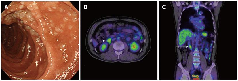
Figure 3 A 60-year-old male patient with duodenal follicular lymphoma without nodal lesions (Lugano system stage I, grade 1).
A: An esophagogastroduodenoscopy detected features typical of primary duodenal follicular lymphoma, including small whitish nodules; B, C: 18F-fluorodeoxyglucose positron emission tomography detected tracer uptake in the duodenal second portion (indicated with arrows).
Figure 4 A 65-year-old female patient with duodenal and jejunal follicular lymphoma and intra-abdominal lymph node involvement (Lugano system stage II-1, grade 1).
A: An esophagogastroduodenoscopy revealed small whitish nodules present in the duodenum; B: Video capsule endoscopy also identified small nodules present in the jejunum; C, D: 18F-fluorodeoxyglucose positron emission tomography detected tracer uptake in the duodenum (C) and the jejunum (D).
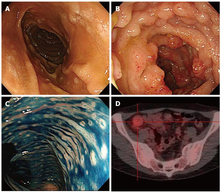
Figure 5 A 37-year-old female patient with systemic follicular lymphoma and extended gastrointestinal involvement from the duodenum to the rectum (stage IV, grade 1).
A: An esophagogastroduodenoscopy detected small whitish nodules present in the duodenum; B, C: A colonoscopy revealed multiple polyps present in the ileum (B), cecum, colon (C), and rectum. Jejunal involvement was confirmed by video capsule endoscopy; D: During 18F-fluorodeoxyglucose positron emission tomography, tracer uptake was only noted in the ileum and colon.
Figure 6 A 61-year-old female patient with follicular lymphoma and duodenal involvement (Lugano system stage IV, grade 1).
A: In the duodenum, small whitish nodules were observed; B: 18F-fluorodeoxyglucose positron emission tomography (18F-FDG-PET) detected tracer uptake in the duodenum (indicated with an arrow); C: 18F-FDG-PET identified diffuse lymphoma infiltration into the spleen (indicated with an arrow), which was not detected by computed tomography scanning. In this case, these results upgraded the clinical stage.
- Citation: Iwamuro M, Okada H, Takata K, Shinagawa K, Fujiki S, Shiode J, Imagawa A, Araki M, Morito T, Nishimura M, Mizuno M, Inaba T, Suzuki S, Kawai Y, Yoshino T, Kawahara Y, Takaki A, Yamamoto K. Diagnostic role of 18F-fluorodeoxyglucose positron emission tomography for follicular lymphoma with gastrointestinal involvement. World J Gastroenterol 2012; 18(44): 6427-6436
- URL: https://www.wjgnet.com/1007-9327/full/v18/i44/6427.htm
- DOI: https://dx.doi.org/10.3748/wjg.v18.i44.6427


