Copyright
©2012 Baishideng Publishing Group Co.
World J Gastroenterol. Nov 7, 2012; 18(41): 5986-5989
Published online Nov 7, 2012. doi: 10.3748/wjg.v18.i41.5986
Published online Nov 7, 2012. doi: 10.3748/wjg.v18.i41.5986
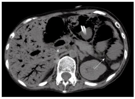
Figure 1 Preoperative computed tomography.
There was a massive amount of portomesenteric venous gas (arrow head) and pneumatosis intestinalis in the small intestine (arrow).
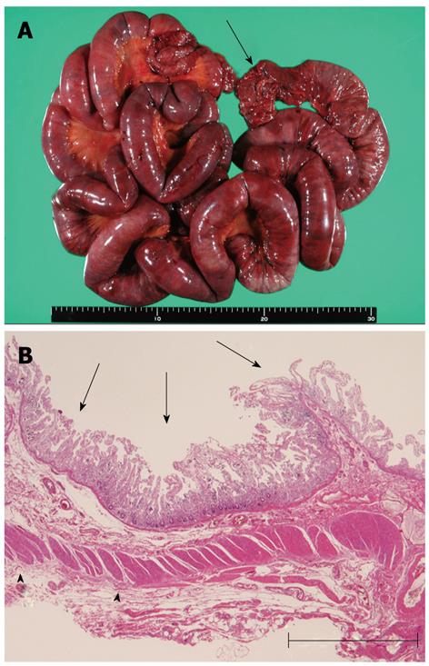
Figure 2 Macroscopic findings of resected tissue and specimen.
A: Macroscopic findings of resected tissue. The specimen measured 150 cm in length. There was diffuse mucosal necrosis in two thirds of oral-sided area, and mottled necrosis in the remaining area. Arrow: Oral stump; B: Microscopic findings of the resected specimen. Coagulation necrosis of the mucosa and lamina propria was observed (arrows). The external longitudinal layer of the muscularis propria was partially diminished (arrow heads). Hematoxylin and eosin, Bar = 500 μm.
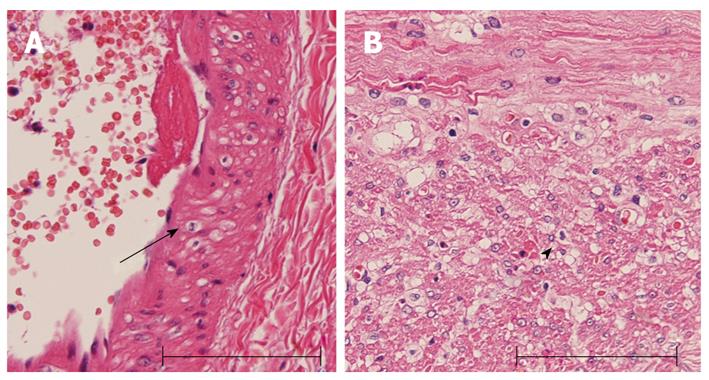
Figure 3 Vacuolar degeneration.
A: Artery wall (arrow); B: Muscular layer (arrow head). Hematoxylin and eosin, Bar = 100 μm.
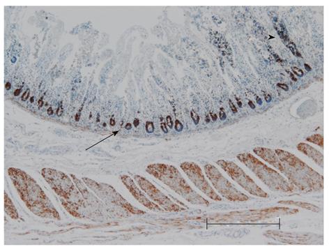
Figure 4 Immunohistochemical study.
Anti-mitochondrial antibody is markedly expressed in the crypts (arrow) and preserved mucosa (arrow head). Bar = 500 μm.
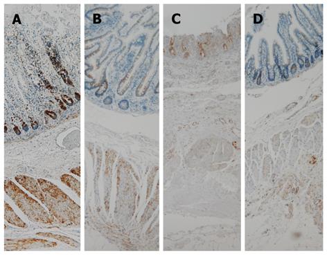
Figure 5 Expression of anti-mitochondrial antibody.
A: Necrotic area of the present patient; B: Non-necrotic area of the present patient; C: Necrotic area of strangulated small intestine of a non-myopathy, encephalopathy, lactic acidosis, and stroke-like episodes (MELAS) patient; D: Non-necrotic c area in the strangulated small intestine of a non-MELAS patient. Bar = 500 μm.
- Citation: Fukuyama K, Ishikawa Y, Ogino T, Inoue H, Yamaoka R, Hirose T, Nishihira T. Mucosal necrosis of the small intestine in myopathy, encephalopathy, lactic acidosis, and stroke-like episodes syndrome. World J Gastroenterol 2012; 18(41): 5986-5989
- URL: https://www.wjgnet.com/1007-9327/full/v18/i41/5986.htm
- DOI: https://dx.doi.org/10.3748/wjg.v18.i41.5986


