Copyright
©2012 Baishideng Publishing Group Co.
World J Gastroenterol. Nov 7, 2012; 18(41): 5982-5985
Published online Nov 7, 2012. doi: 10.3748/wjg.v18.i41.5982
Published online Nov 7, 2012. doi: 10.3748/wjg.v18.i41.5982
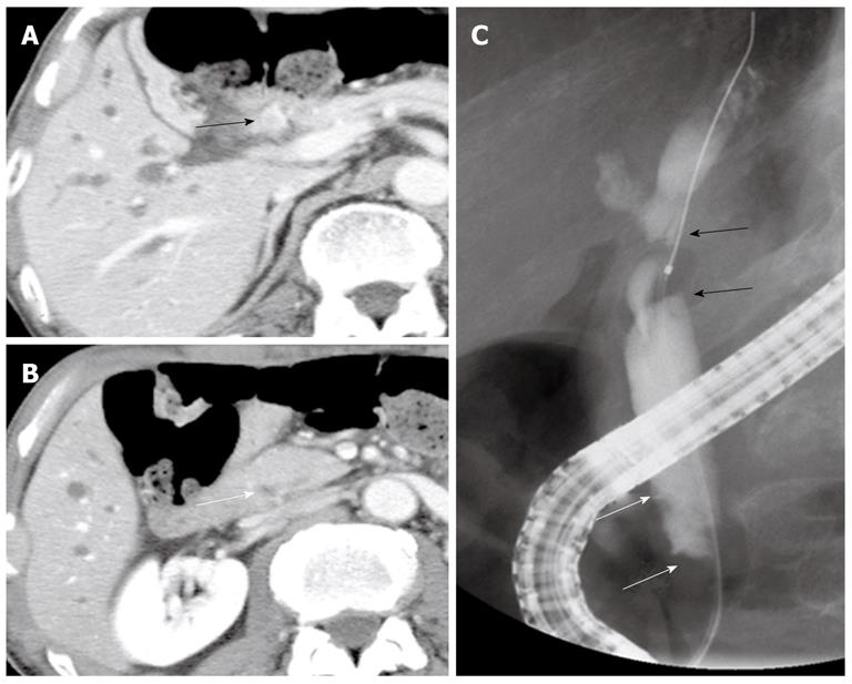
Figure 1 Imaging findings before the first surgery.
A: Computed tomography showed a well enhanced mass in the middle and superior parts of the bile duct (black arrow); B: No mass was detected in the inferior part of the bile duct (white arrow); C: Endoscopic retrograde cholangiopancreatography revealed a tuberous filling defect in the middle and superior parts of the bile duct (black arrows) and mild stenosis in the inferior part (white arrows).
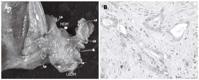
Figure 2 Macroscopic and microscopic findings of resected specimen at the first surgery.
A: Resected specimen of the extra-hepatic bile duct showed whitish tuberous tumor in the middle and inferior parts of the bile duct (cancer marked by the white arrows); B: Microscopically, the tumor was moderately differentiated tubular adenocarcinoma with invasive growth (hematoxylin-eosin; magnification; × 100). GB: Gallbladder; HDM: Hepatic duct margin; LBDM: Lower bile duct margin.
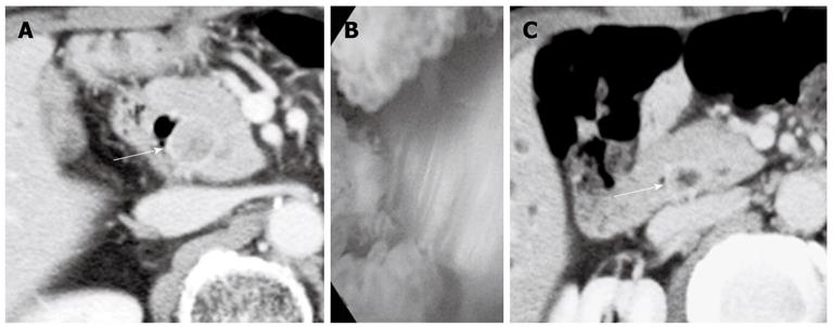
Figure 3 Imaging findings before the second surgery and retrospectively reviewed computed tomography finding before the first surgery.
A: Computed tomography (CT) taken 11 mo after the first surgery showed enhanced inferior bile duct wall (white arrow) and slightly enhanced tumor within the duct; B: Cholangioscopy revealed a papillary tumor in the remaining inferior bile duct; C: Retrospective review of the CT images before the first surgery revealed enhanced inferior bile duct wall (white arrow) only on the delayed phase.
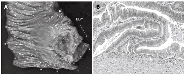
Figure 4 Macroscopic and microscopic findings of resected specimen at the second surgery.
A: Specimen resected during pancreaticoduodenectomy. Note the papillary tumor in the inferior bile duct (white arrow); B: Microscopic examination showed papillary adenocarcinoma with expansive growth (hematoxylin-eosin, magnification; × 100). Tumor cells were confined within the fibromuscular coat. BDM: Bile duct margin.
- Citation: Sumiyoshi T, Shima Y, Kozuki A. Synchronous double cancers of the common bile duct. World J Gastroenterol 2012; 18(41): 5982-5985
- URL: https://www.wjgnet.com/1007-9327/full/v18/i41/5982.htm
- DOI: https://dx.doi.org/10.3748/wjg.v18.i41.5982


