©2012 Baishideng Publishing Group Co.
World J Gastroenterol. Oct 28, 2012; 18(40): 5826-5829
Published online Oct 28, 2012. doi: 10.3748/wjg.v18.i40.5826
Published online Oct 28, 2012. doi: 10.3748/wjg.v18.i40.5826
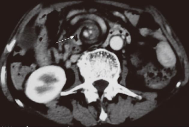
Figure 1 Initial contrast-enhanced computed tomography scan of the abdomen showed mesentery and superior mesenteric artery with "whirlpool" signs (arrow).
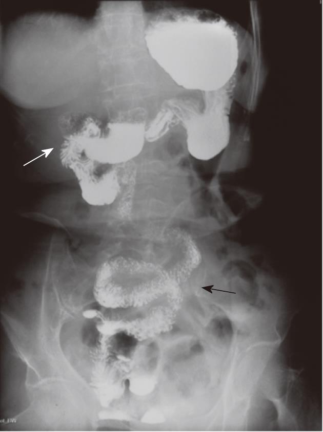
Figure 2 Upper gastrointestinal barium studies demonstrated duodenal diverticulum (white arrow) and "corkscrew" configuration (black arrow) of proximal small bowel.
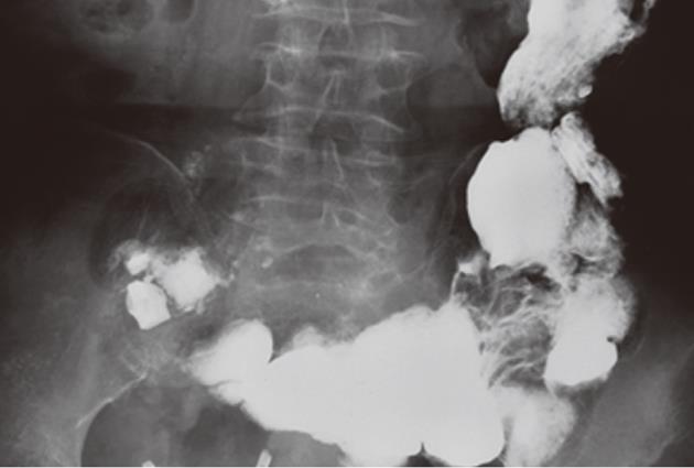
Figure 3 Upper gastrointestinal barium study showed multiple giant jejunal diverticula.
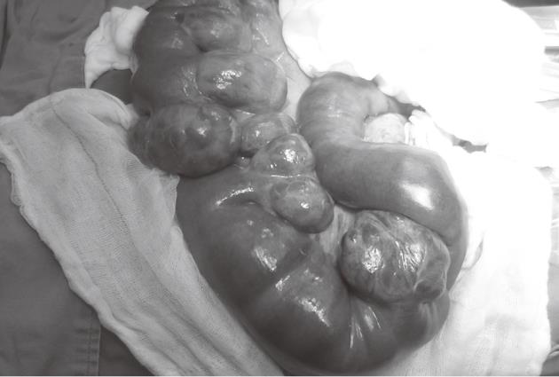
Figure 4 Extremely enlarged jejuna were seen after distortion.
Enlargement starts 20 cm distal to the ligament of Treitz and extends to 120 cm.
- Citation: Hu JL, Chen WZ. Midgut volvulus due to jejunal diverticula: A case report. World J Gastroenterol 2012; 18(40): 5826-5829
- URL: https://www.wjgnet.com/1007-9327/full/v18/i40/5826.htm
- DOI: https://dx.doi.org/10.3748/wjg.v18.i40.5826


