©2012 Baishideng Publishing Group Co.
World J Gastroenterol. Oct 21, 2012; 18(39): 5653-5657
Published online Oct 21, 2012. doi: 10.3748/wjg.v18.i39.5653
Published online Oct 21, 2012. doi: 10.3748/wjg.v18.i39.5653
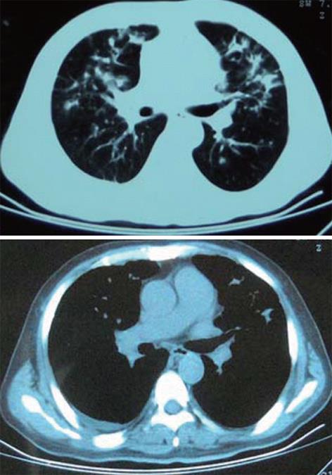
Figure 1 Computed tomography scan of the thorax (January 19, 2009), showing multiple patchy shadows in both lung fields with a little right pleural effusion.
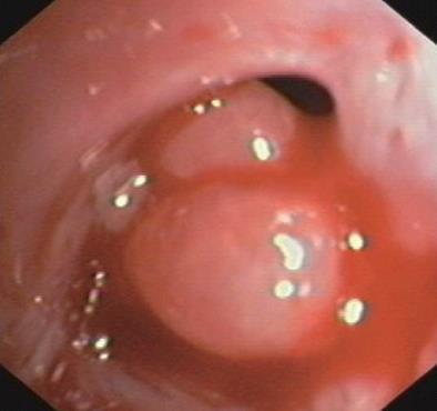
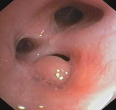
Figure 2 Fibreoptic bronchoscopy (March 9, 2009), showing nodes in the right upper lobe opening.
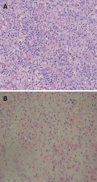
Figure 3 Transfiberoptic bronchoscopy mucosal biopsy, showing polypoid hyperplasia of granulation tissue and hyperplasia of capillary with inflammatory cells (hematoxylin and eosin stain, ×200).
A: Tracheobronchial nodules; B: Left lingular lobe.
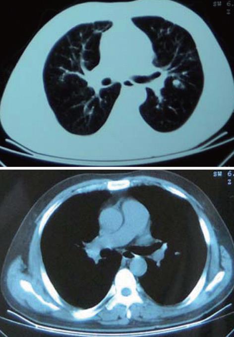
Figure 4 Computed tomography scan of the thorax (April 5, 2009), showing multiple patchy shadows in both lung fields almost disappeared.
Figure 5 Fibreoptic bronchoscopy (April 6, 2009), showing nodes in the right upper lobe opening diminished obviously.
- Citation: Lu DG, Ji XQ, Zhao Q, Zhang CQ, Li ZF. Tracheobronchial nodules and pulmonary infiltrates in a patient with Crohn's disease. World J Gastroenterol 2012; 18(39): 5653-5657
- URL: https://www.wjgnet.com/1007-9327/full/v18/i39/5653.htm
- DOI: https://dx.doi.org/10.3748/wjg.v18.i39.5653


