©2012 Baishideng Publishing Group Co.
World J Gastroenterol. Oct 14, 2012; 18(38): 5485-5488
Published online Oct 14, 2012. doi: 10.3748/wjg.v18.i38.5485
Published online Oct 14, 2012. doi: 10.3748/wjg.v18.i38.5485
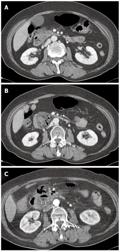
Figure 1 Abdominal computed tomography findings.
A: The pancreas head showed swelling with irregular contour of the pancreatic margin and mild peripancreatic infiltration; B: There is a 5 cm size subtle rim enhancing mass with air bubbles, indicating acute diverticulitis in the second duodenal portion or air forming abscess of the pancreatic head; C: After nine days, a few extraluminal air bubbles (arrow) suspicious for microperforation were found.
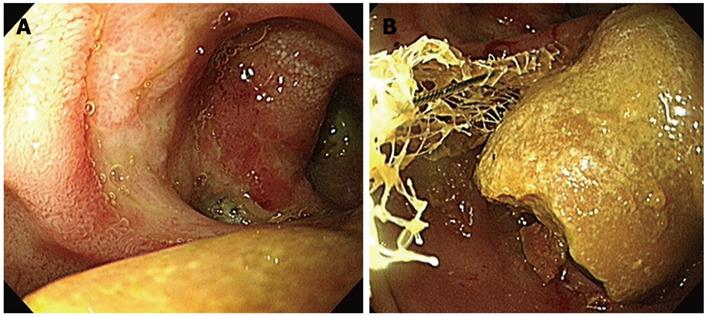
Figure 2 Endoscopic findings.
A: There is a huge luminal obstructing bezoar near the duodenal diverticulum; B: The bezoar was too large to be captured and removed by an endoscopic basket or net.
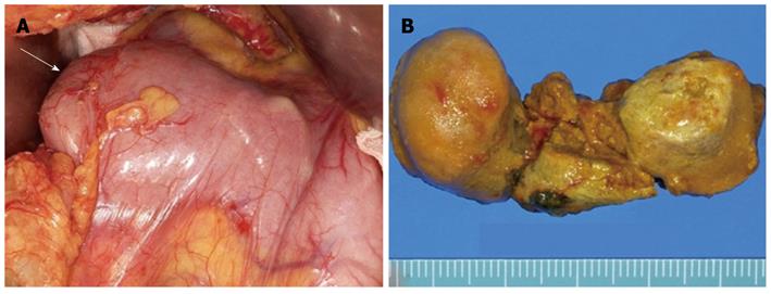
Figure 3 Surgical findings.
A: Obstructed duodenum with impacted huge bezoar (arrow); B: Surgical specimen of the divided bezoar, 5 cm long, 2.5 cm in diameter.
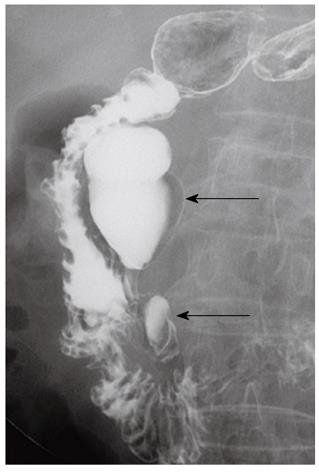
Figure 4 Upper gastrointestinal series show two periampullary diverticula (arrows) in the second duodenal portion.
- Citation: Kim JH, Chang JH, Nam SM, Lee MJ, Maeng IH, Park JY, Im YS, Kim TH, Park IY, Han SW. Duodenal obstruction following acute pancreatitis caused by a large duodenal diverticular bezoar. World J Gastroenterol 2012; 18(38): 5485-5488
- URL: https://www.wjgnet.com/1007-9327/full/v18/i38/5485.htm
- DOI: https://dx.doi.org/10.3748/wjg.v18.i38.5485


