Copyright
©2012 Baishideng Publishing Group Co.
World J Gastroenterol. Sep 21, 2012; 18(35): 4967-4972
Published online Sep 21, 2012. doi: 10.3748/wjg.v18.i35.4967
Published online Sep 21, 2012. doi: 10.3748/wjg.v18.i35.4967
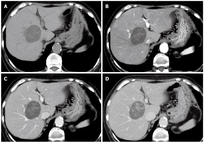
Figure 1 Abdominal computed tomography.
A: Plain; B: Arterial dominant phase; C: Portal venous phase; D: Late venous phase. Abdominal computed tomography (CT) showed an approximately 5-cm well-defined round tumor in segment 8 of the right hepatic lobe. CT showed heterogeneous enhancement in the arterial dominant phase; and delayed the enhancement until the portal venous and late venous phases.
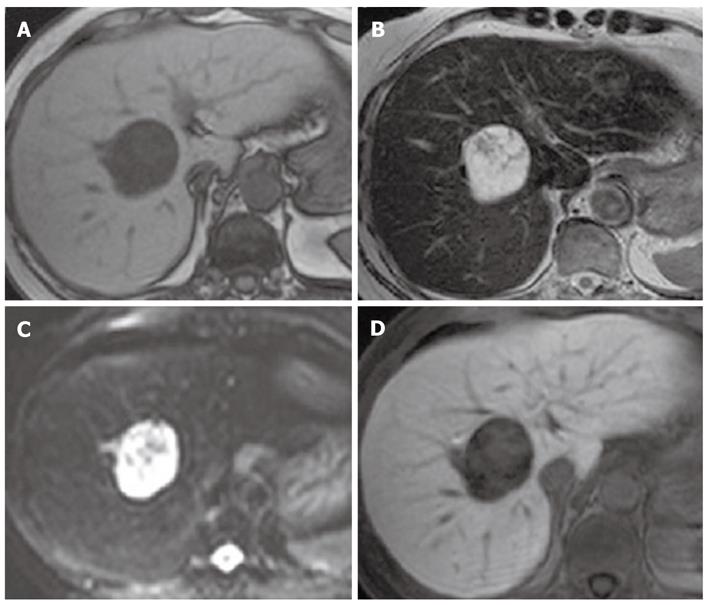
Figure 2 Magnetic resonance imaging.
A: T1-weighted imaging; B: T2-weighted imaging; C: Diffusion-weighted imaging (DWI); D: Hepatobiliary phase. Magnetic resonance imaging (MRI) revealed a hypointense mass on T1-weighted imaging, and a mixed hypo- and hyperintense mass on T2-weighted imaging and on DWI. Gadolinium-ethoxybenzyl-diethylenetriamine penta-acetic acid-enhanced MRI showed a defect in the hepatobiliary phase.
Figure 3 Contrast-enhanced ultrasonography with Sonazoid.
A: B-mode ultrasound; B: Vascular phase; C: Postvascular phase. Ultrasonography showed a 4.6-cm mass that contained multiple hypoechoic cysts with mixed internal septations and solid areas. Contrast-enhanced ultrasonography with Sonazoid showed minute blood flow into the septum and solid areas of the tumor in the vascular phase, and contrast defect of cystic areas (white arrow) and delayed enhancement of solid areas (red arrow) in the postvascular phase.
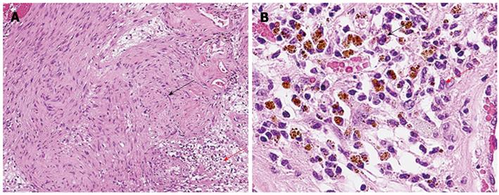
Figure 4 Microscopic examination.
A: B-mode ultrasound. Microscopic examination revealed that Antoni A hypercellular area (black arrow) and Antoni B hypocellular area (red arrow) existed together in a complex; B: Vascular phase. In the Antoni B region, there were edematous stroma, vasodilatation, aggregation of siderophores (black arrow), and slight permeation of chronic inflammatory cells of the histiocyte and lymphocyte types (red arrow).
- Citation: Ota Y, Aso K, Watanabe K, Einama T, Imai K, Karasaki H, Sudo R, Tamaki Y, Okada M, Tokusashi Y, Kono T, Miyokawa N, Haneda M, Taniguchi M, Furukawa H. Hepatic schwannoma: Imaging findings on CT, MRI and contrast-enhanced ultrasonography. World J Gastroenterol 2012; 18(35): 4967-4972
- URL: https://www.wjgnet.com/1007-9327/full/v18/i35/4967.htm
- DOI: https://dx.doi.org/10.3748/wjg.v18.i35.4967


