Copyright
©2012 Baishideng Publishing Group Co.
World J Gastroenterol. Aug 28, 2012; 18(32): 4435-4440
Published online Aug 28, 2012. doi: 10.3748/wjg.v18.i32.4435
Published online Aug 28, 2012. doi: 10.3748/wjg.v18.i32.4435
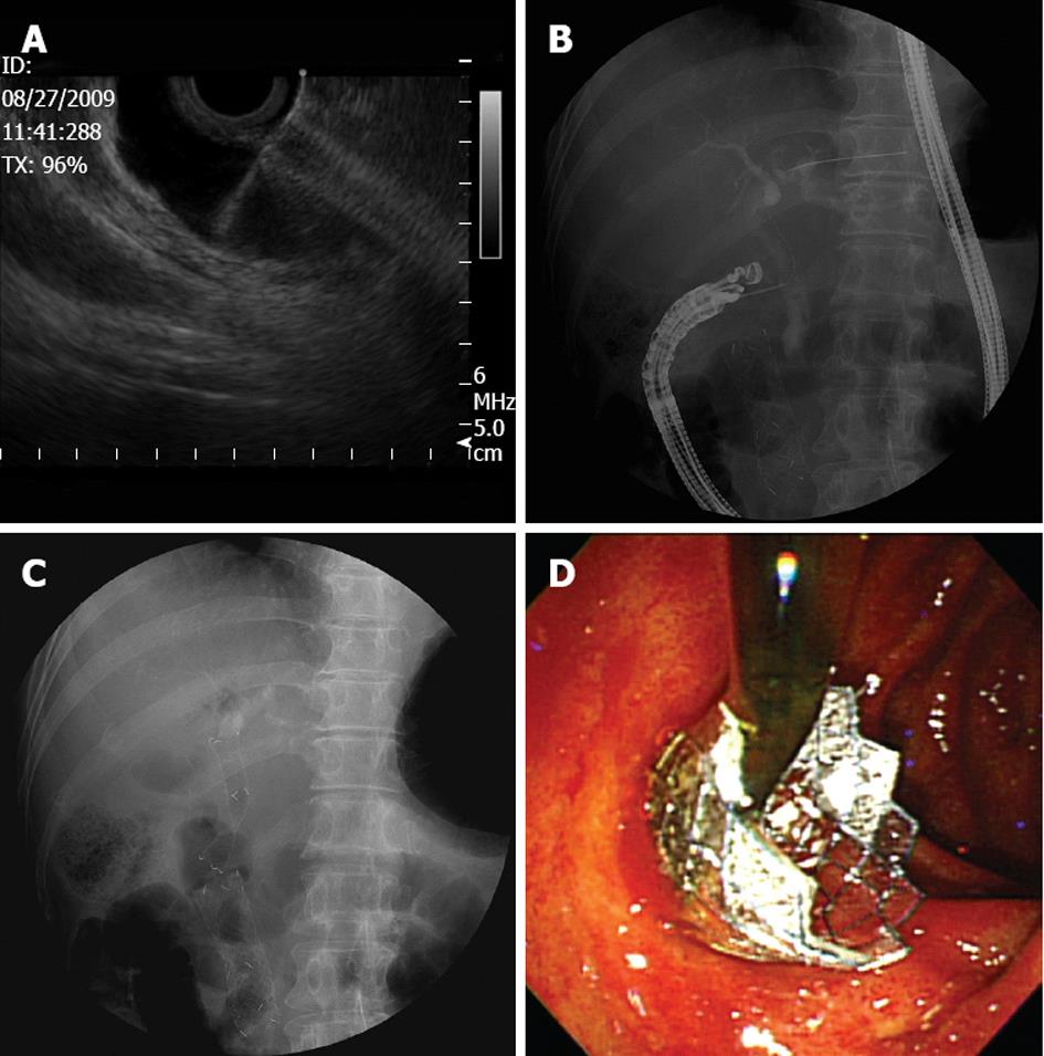
Figure 1 Technique for endoscopic ultrasound-guided choledochoduodenostomy with a fully covered self-expandable metallic stent.
A: The bile duct was punctured with a 19-gauge needle under linear-array echoendoscopic guidance; B: A guidewire was introduced through the needle; C: A fully covered self-expandable metallic stent was inserted under fluoroscopic guidance; D: Illustration of a biliary stent extending from the first portion of the duodenum to the extrahepatic bile duct.
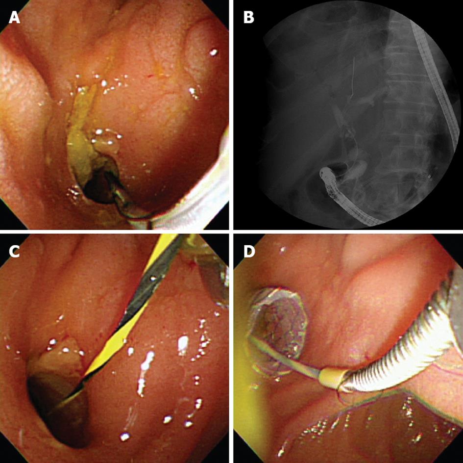
Figure 2 Endoscopic re-intervention for the distal migration of a fully covered self-expandable metallic stent.
A: Endoscopic view showing the patent opening of a fistulous tract between the extrahepatic bile duct and duodenum; B: A fluoroscopic image demonstrated the distal stent migration; C: A guidewire was simply inserted through the existing choledochoduodenal fistula; D: Insertion of a new fully covered self-expandable metallic stent through a mature fistula with a large diameter.
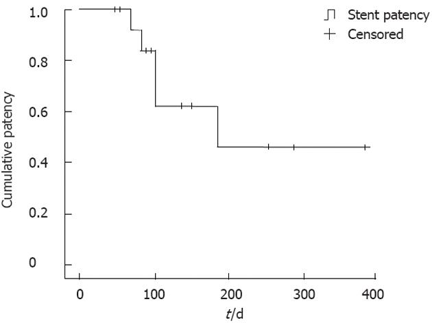
Figure 3 Kaplan-Meier survival curve showing stent patency.
- Citation: Song TJ, Hyun YS, Lee SS, Park DH, Seo DW, Lee SK, Kim MH. Endoscopic ultrasound-guided choledochoduodenostomies with fully covered self-expandable metallic stents. World J Gastroenterol 2012; 18(32): 4435-4440
- URL: https://www.wjgnet.com/1007-9327/full/v18/i32/4435.htm
- DOI: https://dx.doi.org/10.3748/wjg.v18.i32.4435


