©2012 Baishideng Publishing Group Co.
World J Gastroenterol. Jul 28, 2012; 18(28): 3662-3672
Published online Jul 28, 2012. doi: 10.3748/wjg.v18.i28.3662
Published online Jul 28, 2012. doi: 10.3748/wjg.v18.i28.3662
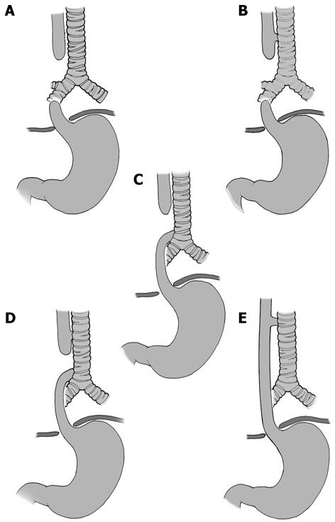
Figure 1 Classification of esophageal atresia/tracheoesophageal fistula.
A: Esophageal atresia (EA) without tracheoesophageal fistula (TEF); B: Proximal TEF with distal EA; C: Distal TEF with proximal EA; D: Proximal and distal TEF; E: TEF without EA or “H”-type TEF.
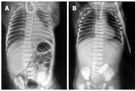
Figure 2 Plain X-rays of the chest and abdomen of two neonates with esophageal atresia.
A: The non-progression of an orogastric catheter in the blind esophageal pouch and the presence of air in the stomach diagnose esophageal atresia with distal tracheoesophageal fistula; B: The radiopaque tube in the blind esophageal pouch and the absence of air in the stomach identify esophageal atresia without tracheoesophageal fistula.
- Citation: Pinheiro PFM, Simões e Silva AC, Pereira RM. Current knowledge on esophageal atresia. World J Gastroenterol 2012; 18(28): 3662-3672
- URL: https://www.wjgnet.com/1007-9327/full/v18/i28/3662.htm
- DOI: https://dx.doi.org/10.3748/wjg.v18.i28.3662


