Copyright
©2012 Baishideng Publishing Group Co.
World J Gastroenterol. Jun 21, 2012; 18(23): 3027-3031
Published online Jun 21, 2012. doi: 10.3748/wjg.v18.i23.3027
Published online Jun 21, 2012. doi: 10.3748/wjg.v18.i23.3027
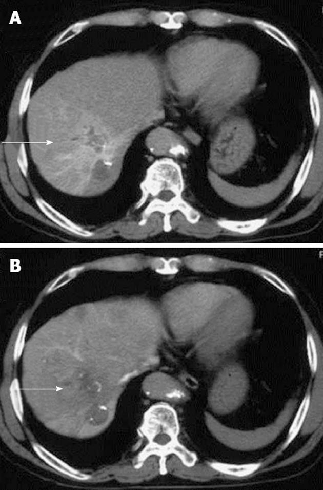
Figure 1 An abdominal computed tomography angiographic scan, showing a diffuse high-density area.
A: An abdominal computed tomography angiographic scan including a lesion previously treated by radiofrequency ablation in segment 8 of the right hepatic lobe, in the arterial phase (arrow); B: The area was washed out on late-phase images (arrow).
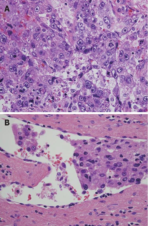
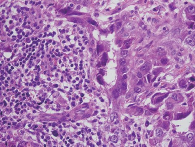
Figure 2 Histopathological examination revealed the presence of considerable necrosis and fibrosis in the tumor.
The hepatocellular carcinoma (HCC) was graded as moderate-grade funicular type. A: Poorly differentiated HCC was present in some regions; B: Peripheral portal vein invasion was detected (hematoxylin and eosin, ×600).
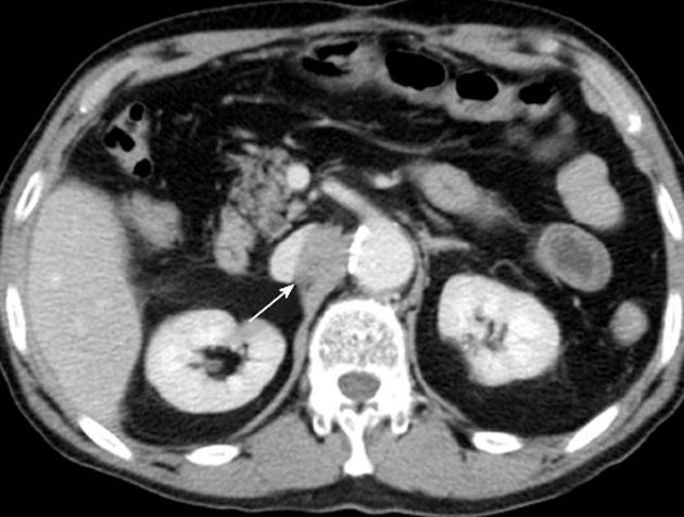
Figure 3 An enhanced computed tomographic scan of the abdomen revealed a tumor (20 mm in diameter, arrow) on the right side of the abdominal aorta.
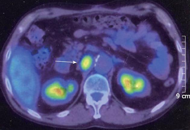
Figure 4 Fluorine-18 fluorodeoxyglucose positron emission tomography revealed that the tumor had an elevated standard uptake value (arrow).
Figure 5 Histopathological examination revealed that the tumor consisted of a broad region of necrosis with poorly differentiated hepatocellular carcinoma on its surface (hematoxylin and eosin, ×600).
- Citation: Ueda J, Yoshida H, Mamada Y, Taniai N, Mineta S, Yoshioka M, Kawano Y, Shimizu T, Hara E, Kawamoto C, Kaneko K, Uchida E. Surgical resection of a solitary para-aortic lymph node metastasis from hepatocellular carcinoma. World J Gastroenterol 2012; 18(23): 3027-3031
- URL: https://www.wjgnet.com/1007-9327/full/v18/i23/3027.htm
- DOI: https://dx.doi.org/10.3748/wjg.v18.i23.3027


