Copyright
©2012 Baishideng Publishing Group Co.
World J Gastroenterol. Jun 14, 2012; 18(22): 2837-2843
Published online Jun 14, 2012. doi: 10.3748/wjg.v18.i22.2837
Published online Jun 14, 2012. doi: 10.3748/wjg.v18.i22.2837
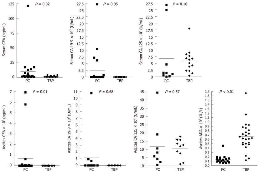
Figure 1 Scatter plots shows the distributions of tumor markers and adenosine deaminase in serum and ascites between peritoneal carcinomatosis group and tuberculous peritonitis group.
All tests were performed by Mann-Whitney U-test. PC: Peritoneal carcinomatosis; TBP: Tuberculous peritonitis; ADA: Adenosine deaminase; CEA: Carcinoembryonic antigen; CA 19-9: Carbohydrate antigen 19-9; CA 125: Carbohydrate antigen 125.
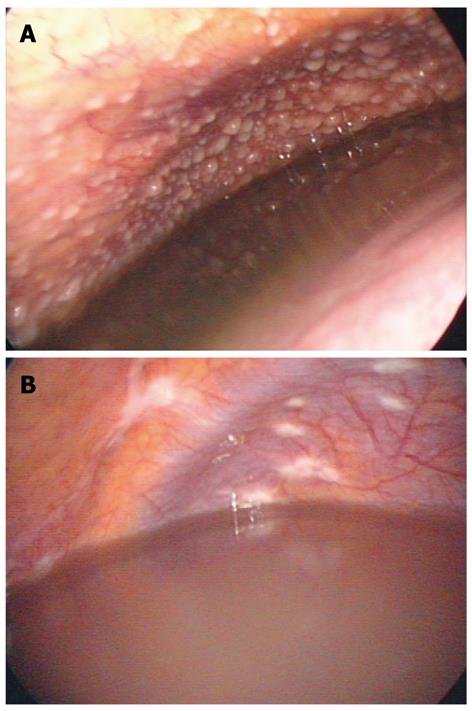
Figure 2 Peritoneoscopic pictures of tuberculous peritonitis and peritoneal carcinomatosis.
A: Peritoneoscopic picture of tuberculous peritonitis of female patient. Multitudinous miliary nodules are seen on the parietal peritoneum. Biopsy revealed caseous granulomatous inflammation and the biopsy specimen stained positive for acid-fast bacilli; B: Colon cancer with peritoneal seeding. Multiple irregular whitish patch lesions were found on the parietal peritoneum. Poorly differentiated adenocarcinoma was documented on biopsy.
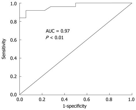
Figure 3 Receiver operating characteristic curve of ascites adenosine deaminase for differentiating between tuberculous peritonitis and peritoneal carcinomatosis.
AUC of this receiver operating characteristic curve is 0.97 (95% CI: 0.92-1.00, P < 0.01). CI: Confidence interval; AUC: Area under the curve.
- Citation: Kang SJ, Kim JW, Baek JH, Kim SH, Kim BG, Lee KL, Jeong JB, Jung YJ, Kim JS, Jung HC, Song IS. Role of ascites adenosine deaminase in differentiating between tuberculous peritonitis and peritoneal carcinomatosis. World J Gastroenterol 2012; 18(22): 2837-2843
- URL: https://www.wjgnet.com/1007-9327/full/v18/i22/2837.htm
- DOI: https://dx.doi.org/10.3748/wjg.v18.i22.2837


