Copyright
©2012 Baishideng Publishing Group Co.
World J Gastroenterol. May 14, 2012; 18(18): 2291-2294
Published online May 14, 2012. doi: 10.3748/wjg.v18.i18.2291
Published online May 14, 2012. doi: 10.3748/wjg.v18.i18.2291
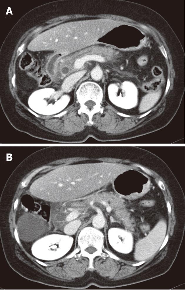
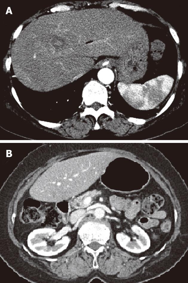
Figure 1 Abdominal computed tomography scans showed diffuse swelling and infiltration in head, body (A) and tail (B) of pancreas without obvious stone, indicating acute pancreatitis.
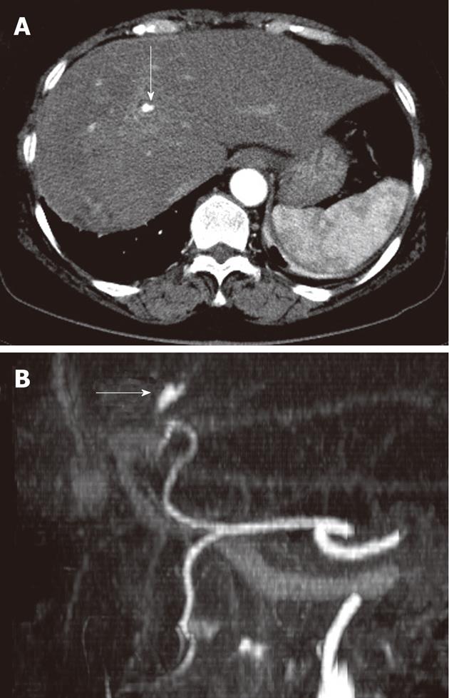
Figure 2 Follow up of abdominal computed tomography scan (A) due to abruptly aggravated abdominal pain and reconstructed computed tomography angiogram (B) showed newly developed hepatic artery pseudoaneurysm in the left lateral segment of liver (white arrows).
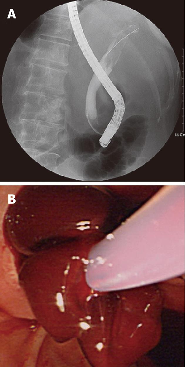
Figure 3 Cholangiogram by endoscopic retrograde cholangiopancreatography showed several amorphous filling defects in the common bile duct (A) and lots of blood clots were seen and removed by basket after endoscopic sphincterotomy (B).
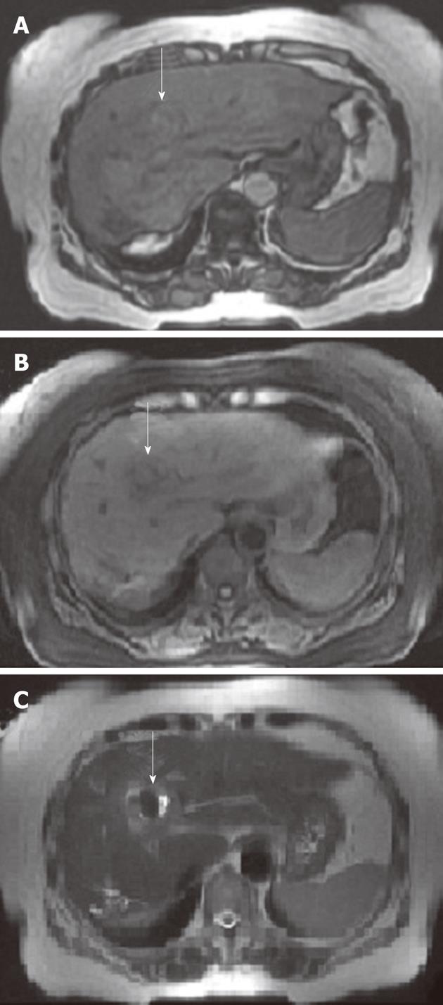
Figure 4 Magnetic resonance imaging scans of the abdomen revealed that hepatic artery pseudoaneurysm was replaced by thrombus formation (white arrows) in the left lateral segment in-phase (A) and out-of-phase (B) T1-weighted images and T2-weighted image (C).
Figure 5 Follow-up of abdominal computed tomography scans 3 mo later showed that both previously noted hepatic artery pseudoaneurysm in the left lateral segment (A) and diffuse swelling and infiltration of pancreas (B) had disappeared.
- Citation: Yu YH, Sohn JH, Kim TY, Jeong JY, Han DS, Jeon YC, Kim MY. Hepatic artery pseudoaneurysm caused by acute idiopathic pancreatitis. World J Gastroenterol 2012; 18(18): 2291-2294
- URL: https://www.wjgnet.com/1007-9327/full/v18/i18/2291.htm
- DOI: https://dx.doi.org/10.3748/wjg.v18.i18.2291


