©2012 Baishideng Publishing Group Co.
World J Gastroenterol. Apr 28, 2012; 18(16): 1940-1945
Published online Apr 28, 2012. doi: 10.3748/wjg.v18.i16.1940
Published online Apr 28, 2012. doi: 10.3748/wjg.v18.i16.1940
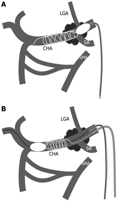
Figure 1 Schematic drawing of microcoil embolization under proximal balloon inflation (A) and distal balloon inflation (B) in the common hepatic artery.
LGA: Left gastric artery; CHA: Common hepatic artery; SA: Splenic artery; SMA: Superior mesenteric artery.
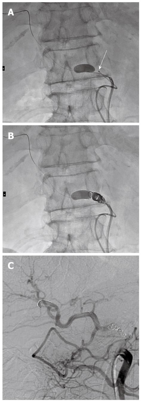
Figure 2 Microcoil embolization under distal balloon inflation.
A: Radiograph during catheterization shows microcatheter (arrow) insertion to the common hepatic artery (CHA) via the celiac artery under distal microballoon inflation in the CHA; B: Radiograph during microcoil embolization shows a tight widthwise frame; C: Superior mesenteric arteriography after microcoil embolization shows blood flow from the superior mesenteric artery to the proper hepatic artery via the pancreatico-duodenal arcades.
Figure 3 Distal migration of the microcoils and the successful withdraw.
A: Following celiac arteriography; B: Microcoil embolization under proximal balloon inflation (arrow) was performed; C: Microcoil migration from the common hepatic artery (CHA) to the proper hepatic artery occurred after deflation of the proximal balloon catheter; D: Under fluoroscopic guidance using the tube angle that enabled the best visualization of the CHA and with the assistance of a microwire, a microballoon catheter was then inserted through the migrated coil and inflated; E: The migrated coils were withdrawn to their original position in the CHA by the inflated balloon catheter.
- Citation: Takasaka I, Kawai N, Sato M, Tanihata H, Sonomura T, Minamiguchi H, Nakai M, Ikoma A, Nakata K, Sanda H. Preoperative microcoil embolization of the common hepatic artery for pancreatic body cancer. World J Gastroenterol 2012; 18(16): 1940-1945
- URL: https://www.wjgnet.com/1007-9327/full/v18/i16/1940.htm
- DOI: https://dx.doi.org/10.3748/wjg.v18.i16.1940


