Copyright
©2011 Baishideng Publishing Group Co.
World J Gastroenterol. Mar 7, 2011; 17(9): 1219-1226
Published online Mar 7, 2011. doi: 10.3748/wjg.v17.i9.1219
Published online Mar 7, 2011. doi: 10.3748/wjg.v17.i9.1219
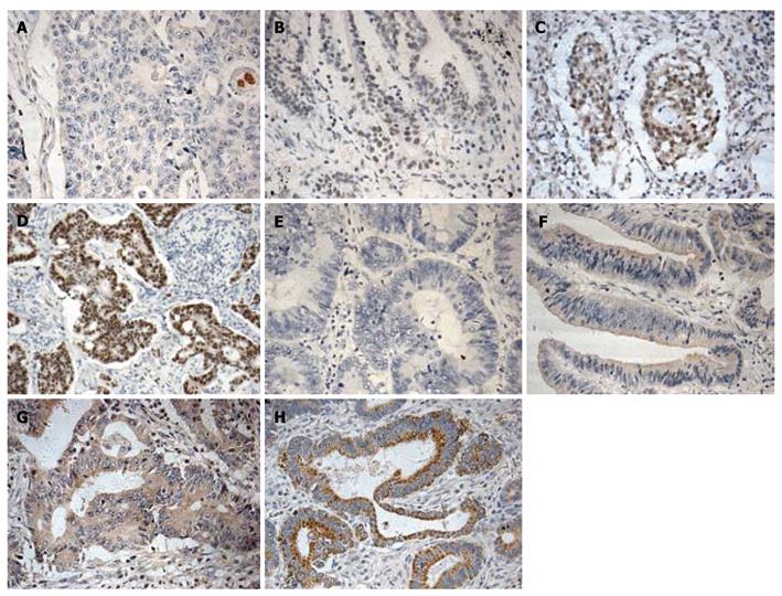
Figure 1 Immunohistochemical labeling for metastasis-associated protein 1, vascular endothelial growth factor-C in colorectal cancer.
Metastasis-associated protein 1 (MTA1) (A-D) and VEGF-C (E-H) expressions are indicated as score 0 (A and E), score 1 (B and F), score 2 (C and G), and score 3 (D and H), respectively. MTA1 showing extremely weak staining of cytoplasm and intense staining of nuclei, while VEGF-C showing staining of cytoplasm of tumor cells (×200).
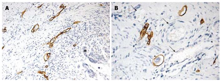
Figure 2 Morphological features of D2-40 positive lymphatic vessels in colorectal cancer.
A: Positive D2-40 stained lymphatic vessels with thin walls and irregular shapes in peritumoral area (asterisk, × 100); B: Positive D2-40 stained lymphatic vessel containing tumor emboli within tumor mass (arrow head) and D2-40-negative erythrocytes (black arrows, × 200).
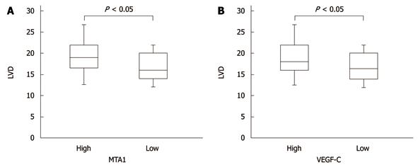
Figure 3 Correlation between metastasis-associated protein 1 or vascular endothelial growth factor-C expression and lymphovascular density.
Analysis of subgroups with low and high metastasis-associated protein 1 (MTA1) and VEGF-C are presented by box bars. Tumors with higher MTA1 and VEGF-C expression showed significantly higher microvessel density than tumors with lower expression. LVD: Lymphovascular density. VEGF-C: Vascular endothelial growth factor-C.
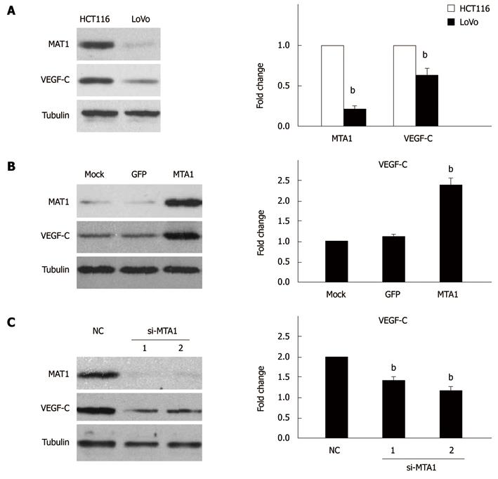
Figure 4 Metastasis-associated protein 1 regulates the expression of vascular endothelial growth factor-C in colorectal cancer cell lines.
Western blotting (left panel) and real-time PCR (right panel) showing different expressions of metastasis-associated protein 1 (MTA1) and VEGF-C in HCT116 and LoVo cell lines (A), increased VEGF-C expression in LoVo cells due to overexpression of MTA1 (B), and decreased VEGF-C expression in HCT116 cells due to knocked down MTA1 (C). 1 and 2 indicate two different si-MTA1 oligonucleotide as showed in "Materials and Methods". All data represent three independent experiments ± SE, with n = 3 and bP < 0.01. VEGF-C: Vascular endothelial growth factor-C.
- Citation: Du B, Yang ZY, Zhong XY, Fang M, Yan YR, Qi GL, Pan YL, Zhou XL. Metastasis-associated protein 1 induces VEGF-C and facilitates lymphangiogenesis in colorectal cancer. World J Gastroenterol 2011; 17(9): 1219-1226
- URL: https://www.wjgnet.com/1007-9327/full/v17/i9/1219.htm
- DOI: https://dx.doi.org/10.3748/wjg.v17.i9.1219


