Copyright
©2011 Baishideng Publishing Group Co.
World J Gastroenterol. Mar 7, 2011; 17(9): 1185-1191
Published online Mar 7, 2011. doi: 10.3748/wjg.v17.i9.1185
Published online Mar 7, 2011. doi: 10.3748/wjg.v17.i9.1185
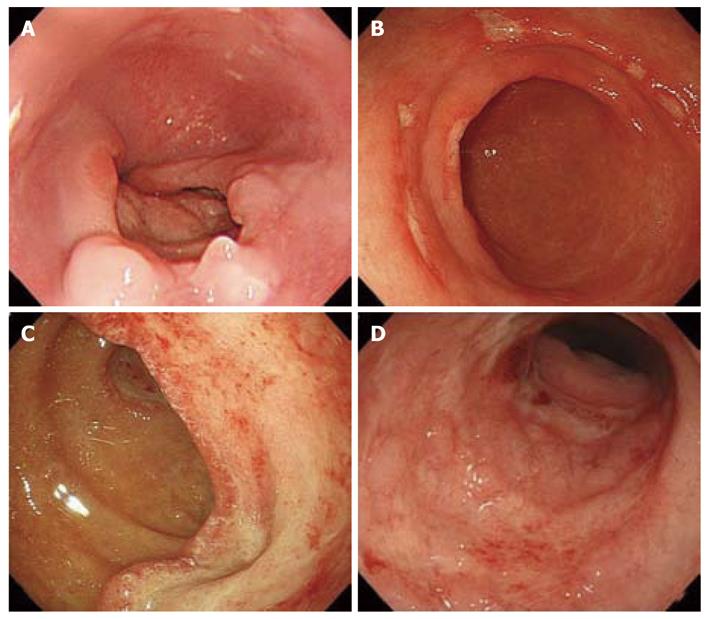
Figure 1 Endoscopic features in cytomegalovirus gastrointestinal disease.
A: Deep, punched-out ulcer in the esophagus; B: Multiple, shallow ulcers in the gastric antrum; C: Large, deep ulcer in the duodenum; D: Multiple erosions and edematous mucosa with ulcer in the sigmoid colon.
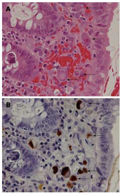
Figure 2 Pathological features in cytomegalovirus gastrointestinal disease.
A: Large cells with intranuclear inclusions or associated with granular cytoplasmic inclusions (hematoxylin and eosin stain); B: Cytomegalovirus (CMV)-infected cells (arrows) show brown coloration in both nuclei and cytoplasm (immunohistochemical staining with anti-CMV).
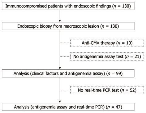
Figure 3 Study design.
CMV: Cytomegalovirus; PCR: Polymerase chain reaction.
- Citation: Nagata N, Kobayakawa M, Shimbo T, Hoshimoto K, Yada T, Gotoda T, Akiyama J, Oka S, Uemura N. Diagnostic value of antigenemia assay for cytomegalovirus gastrointestinal disease in immunocompromised patients. World J Gastroenterol 2011; 17(9): 1185-1191
- URL: https://www.wjgnet.com/1007-9327/full/v17/i9/1185.htm
- DOI: https://dx.doi.org/10.3748/wjg.v17.i9.1185


