Copyright
©2011 Baishideng Publishing Group Co.
World J Gastroenterol. Nov 28, 2011; 17(44): 4867-4874
Published online Nov 28, 2011. doi: 10.3748/wjg.v17.i44.4867
Published online Nov 28, 2011. doi: 10.3748/wjg.v17.i44.4867
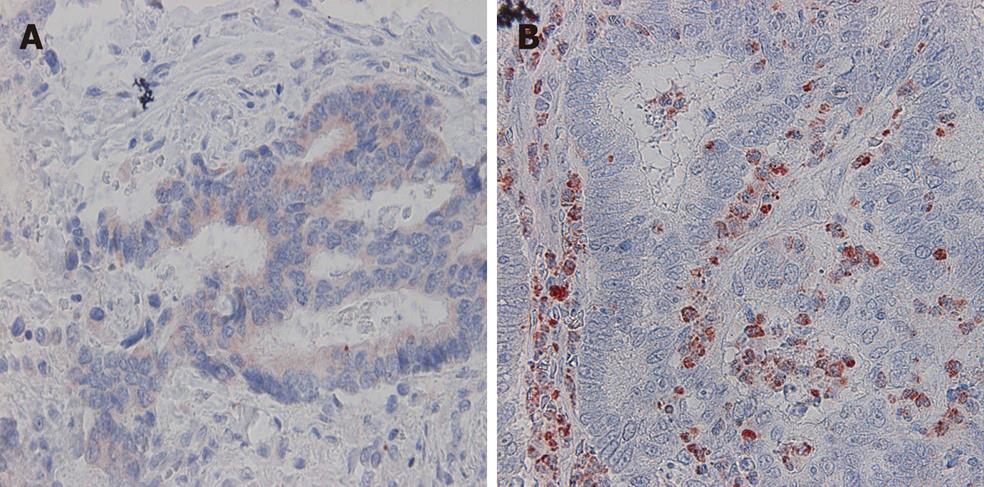
Figure 1 Immunohistochemical of colorectal cancer tissues used the anti-vascular endothelial growth factor-A antibody.
A: Vascular endothelial growth factor (VEGF)-A was expressed in tumor cells but not in stromal cells; B: VEGF-A was expressed in stromal cells but not in tumor cells.
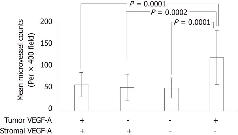
Figure 2 Microvessel density of vascular endothelial growth factor-A expression status.
In s-vascular endothelial growth factor (VAEG)-A positive cases, microvessel density (MVD) was maintained at a low score regardless tumor VEGF-A (t-VEGF-A) expression. In s-VEGF-A negative cases, MVD was influenced by t-VEGF-A expression.
Figure 3 Disease-free survival of patients with stagesII and III colorectal cancer.
A: t-vascular endothelial growth factor (VAGE)-A positive vs negative. The log-rank test indicates P= 0.24 (not significant); B: s-VEGF-A positive vs negative. The log-rank test statistical analysis indicates a significant difference (P = 0.0005). VEGF: Vascular endothelial growth factor.
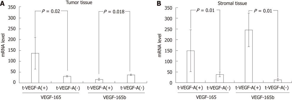
Figure 4 mRNA level of VEGF165 and VEGF165b semi-quantified by real-time polymerase chain reaction in tumor and stromal tissues.
A: In tumor tissue, only vascular endothelial growth factor (VEGF) 165 expressed in t-VEGF-A positive cases; B: In stromal tissues, both VEGF165 and VEGF165b expressed in s-VEGF-A positive case.
Figure 5 Correlations of vascular endothelial growth factor 165b expression in stromal tissue and tumors with venous and lymphatic invasion.
A: Vascular endothelial growth factor (VEGF) 165b mRNA level in v0 cases was significantly higher than those in v1 cases; B: There were no significant differences of VEGF165bmRNA levels among degrees of the lymphatic invasion. NS: Not significant.
Figure 6 Relationship between the level of vascular endothelial growth factor 165 mRNA in tumor, vascular endothelial growth factor 165b mRNA in stromal tissues and microvessel density.
Twenty cases are arrayed on the X axis in ascending order of the amount of vascular endothelial growth factor (VEGF)165b expression. A: The score of microvessel density (MVD); B: The mRNA level of VEGF165 and VEGF165b. MVD was maintained at a low level in the cases in which VEGF165b expressed in stromal tissues.
- Citation: Tayama M, Furuhata T, Inafuku Y, Okita K, Nishidate T, Mizuguchi T, Kimura Y, Hirata K. Vascular endothelial growth factor 165b expression in stromal cells and colorectal cancer. World J Gastroenterol 2011; 17(44): 4867-4874
- URL: https://www.wjgnet.com/1007-9327/full/v17/i44/4867.htm
- DOI: https://dx.doi.org/10.3748/wjg.v17.i44.4867


