Copyright
©2011 Baishideng Publishing Group Co.
World J Gastroenterol. Nov 28, 2011; 17(44): 4839-4844
Published online Nov 28, 2011. doi: 10.3748/wjg.v17.i44.4839
Published online Nov 28, 2011. doi: 10.3748/wjg.v17.i44.4839
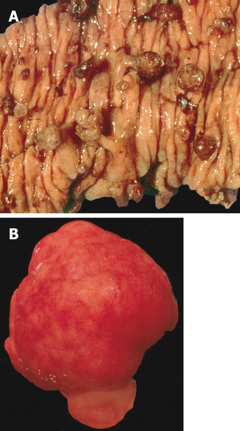
Figure 1 Macroscopic appearance of juvenile polyposis.
A: Bowel resection of a patient with juvenile polyposis syndrome showing multiple spherical pedunculated polyps with a smooth surfaces; B: Gross appearance of a juvenile polyp from a patient with juvenile polyposis syndrome. Note the smooth surface, in contrast with a Peutz-Jeghers polyp.
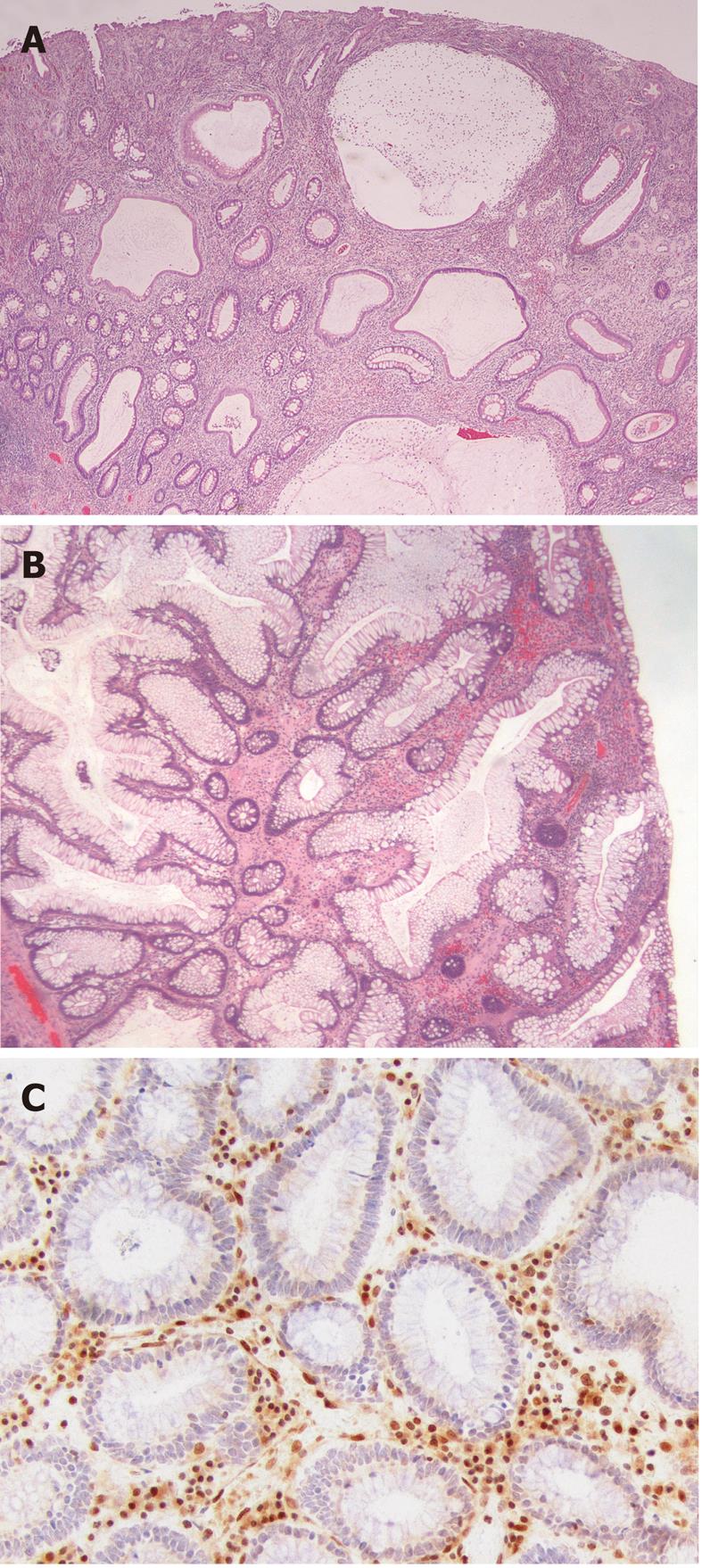
Figure 2 Histological appearance of juvenile polyposis.
A: Histological section of a juvenile polyp from a juvenile polyposis patient with a germline mutation of BMPR1A. Typically, juvenile polyps are characterized by prominent lamina propria with edema and inflammatory cells, and cystically dilated glands lined by cuboidal to columnar epithelium with reactive changes; B: Histological section of a juvenile polyp from a juvenile polyposis patient with a germline mutation of SMAD4. This polyp shows relatively fewer stroma, fewer dilated glands and more proliferative smaller glands; C: SMAD4 immunohistochemistry on a juvenile polyp showing absent SMAD4 expression in the epithelium, indicating that this patient carries a germline SMAD4 mutation.
- Citation: Brosens LA, Langeveld D, Hattem WAV, Giardiello FM, Offerhaus GJA. Juvenile polyposis syndrome. World J Gastroenterol 2011; 17(44): 4839-4844
- URL: https://www.wjgnet.com/1007-9327/full/v17/i44/4839.htm
- DOI: https://dx.doi.org/10.3748/wjg.v17.i44.4839


