©2011 Baishideng Publishing Group Co.
World J Gastroenterol. Nov 21, 2011; 17(43): 4757-4771
Published online Nov 21, 2011. doi: 10.3748/wjg.v17.i43.4757
Published online Nov 21, 2011. doi: 10.3748/wjg.v17.i43.4757
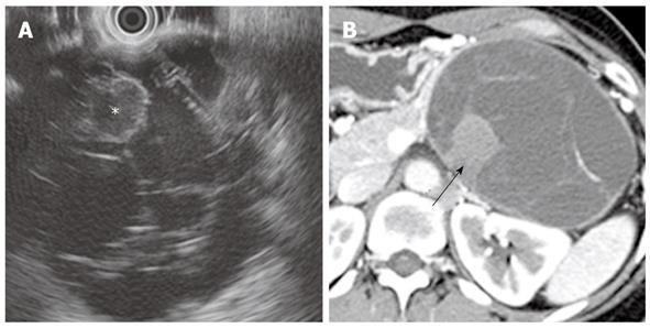
Figure 1 Mucinous cystadenoma of the pancreas in a 40-year-old woman.
A: Endoscopic ultrasound image shows a complex multiloculated cystic mass in the pancreatic tail. Note a hyperechoic locule (asterisk) within the cystic tumor; B: Contrast-enhanced CT image shows a well-circumscribed multiloculated mass in the tail of the pancreas with enhancement of thin internal septa and the peripheral wall. Note a highly attenuated locule (arrow) within the cystic tumor. CT: Computed tomography.
Figure 2 Mucinous cystadenocarcinoma of the pancreas in a 44-year-old woman.
A, B: Non-enhanced (A) and contrast-enhanced (B) CT images show a well-circumscribed multi-septated cystic mass with enhancing soft tissue components (arrowheads) in the tail of the pancreas; C: T2-weighted MR image shows a large multilocular cystic mass with high signal intensity and peripheral soft tissue components with low signal intensity (arrowheads) in the pancreatic tail. CT: Computed tomography; MR: Magnetic resonance.
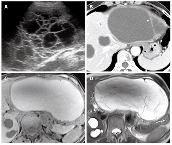
Figure 3 Biliary cystadenoma in the liver of a 55-year-old woman.
A: Ultrasound image shows a multiseptated cystic mass in the liver; B: Contrast-enhanced CT image shows a well-circumscribed cystic liver mass, which has similar attenuation to that of hepatic cysts. Note the internal septa with nodular thickening (arrowhead); C, D: T1- (C) and T2-weighted (D) MR images show a multiseptated cystic mass with hyperintensity. CT: Computed tomography; MR: Magnetic resonance.
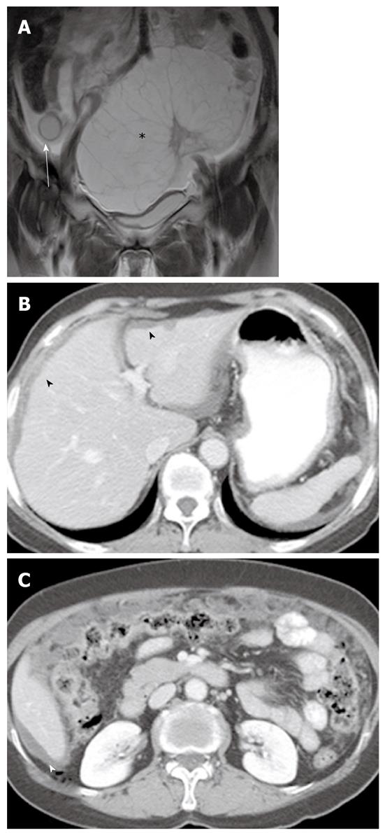
Figure 4 Coexistence of a mucinous cystadenoma of the appendix and ovary in a 63-year-old woman.
A: Coronal T2-weighted MR image shows a well-defined cystic mass with hyperintensity in the appendix (arrow) and a multiloculated cystic mass in the left ovary (asterisk); B, C: Contrast-enhanced CT images show low-attenuation mucinous deposits in the peritoneal cavity and scalloping of the liver margin (arrowheads), suggestive of pseudomyxoma peritonei. CT: Computed tomography; MR: Magnetic resonance.
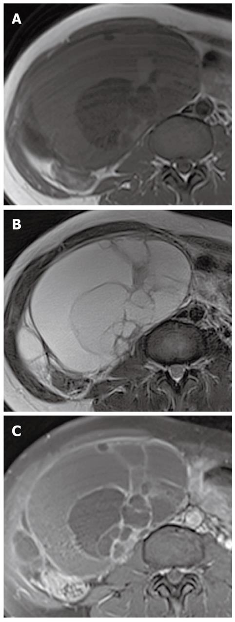
Figure 5 Mucinous borderline tumor of the right ovary in a 41-year-old woman.
A, B: T1- (A) and T2-weighted (B) MR images show a cystic mass with large numbers of loculi with varying signal intensity in the right ovary; C: Contrast-enhanced fat-saturated T1-weighted MR image shows the enhancement of multiple internal septa and wall in the cystic mass. MR: Magnetic resonance.
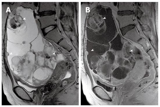
Figure 6 Mucinous cystadenocarcinoma of the left ovary in a 47-year-old woman.
A: Sagittal T2-weighted MR image shows a huge multilocular ovarian cystic mass with multiple hypointense solid components (arrowheads); B: Sagittal contrast-enhanced T1-weighted MR image shows an ovarian cystic mass with enhancement of internal septa and solid components (arrowheads). MR: Magnetic resonance.
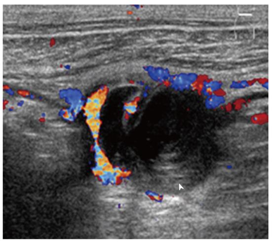
Figure 7 Mucocele of the appendix in a 54-year-old man.
Ultrasound image shows an anechoic round mass in the appendix with echogenic layers (arrowhead) (the so-called “onion-skin” sign).
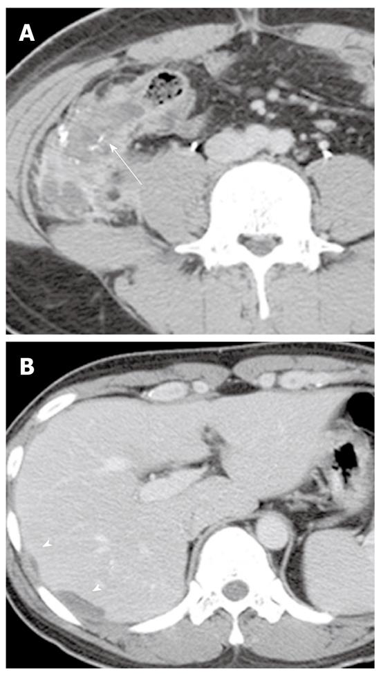
Figure 8 Mucinous cystadenocarcinoma of the appendix in a 26-year-old man.
A: Contrast-enhanced CT image shows a complex hypoattenuating mass with enhancing solid portions and punctate calcifications in the appendix (arrow); B: Contrast-enhanced CT image of the upper abdomen shows loculations of fluid scalloping on the liver surface (arrowheads), providing evidence of a mass effect. CT: Computed tomography.
Figure 9 Intraductal papillary mucinous neoplasm of the pancreas in an 80-year-old woman.
A: Coronal T2-weighted RARE MR image shows cord-like hypointense mucin in the dilated main pancreatic duct (arrow). Note multiple stones in the dilated common bile duct; B: Axial contrast-enhanced fat-saturated T1-weighted MR image demonstrates a fistula (asterisk) between the dilated main pancreatic duct and the stomach. Note mural nodules (arrowheads) within the dilated main pancreatic duct, which strongly suggest a malignant Intraductal papillary mucinous neoplasm; C: Endoscopy image reveals mucin leaking from the papilla. RARE: True rapid acquisition with relaxation enhancement; MR: Magnetic resonance.
Figure 10 Intraductal papillary mucinous neoplasm of the bile duct in a 58-year-old woman.
A: Contrast-enhanced CT image shows severe dilatation of the left intrahepatic ducts without visible intraductal mass. Note a small stone in the dilated intrahepatic duct (arrowhead); B: Coronal T2-weighted RARE MR image shows dilatation of the intrahepatic and extrahepatic ducts. Note the hypointense intraductal mass (arrow) in the left intrahepatic duct; C: Endoscopic retrograde cholangiogram image shows a filling defect (arrow) within the marked dilated extrahepatic bile duct, due to mucin; D: Coronal gadoxetic acid-enhanced T1-weighted MR image obtained at 60 min post-injection shows contrast-filled bile duct with an elongated, low signal intensity lesion (asterisk), which represents mucin; E: Endoscopy image reveals mucin leaking from the papilla. CT: Computed tomography; RARE: True rapid acquisition with relaxation enhancement; MR: Magnetic resonance.
Figure 11 Signet ring cell carcinoma of the stomach in a 45-year-old woman.
Coronal reformatted contrast-enhanced CT image shows diffuse gastric wall thickening with strong enhancement (arrowhead) along the lesser and greater curvature of the stomach. CT: Computed tomography.
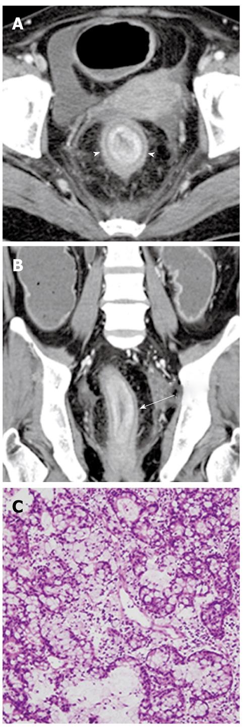
Figure 12 Signet ring cell carcinoma of the rectum in a 42-year-old man.
A, B: Axial (A) and coronal reformatted (B) contrast-enhanced CT images show concentric wall thickening with malignant target sign (arrowheads in A, arrow in B) in the rectum; C: Photomicrograph image (original magnification, × 200; HE stain) shows multiple signet ring cells. CT: Computed tomography; HE: Hematoxylin and eosin.
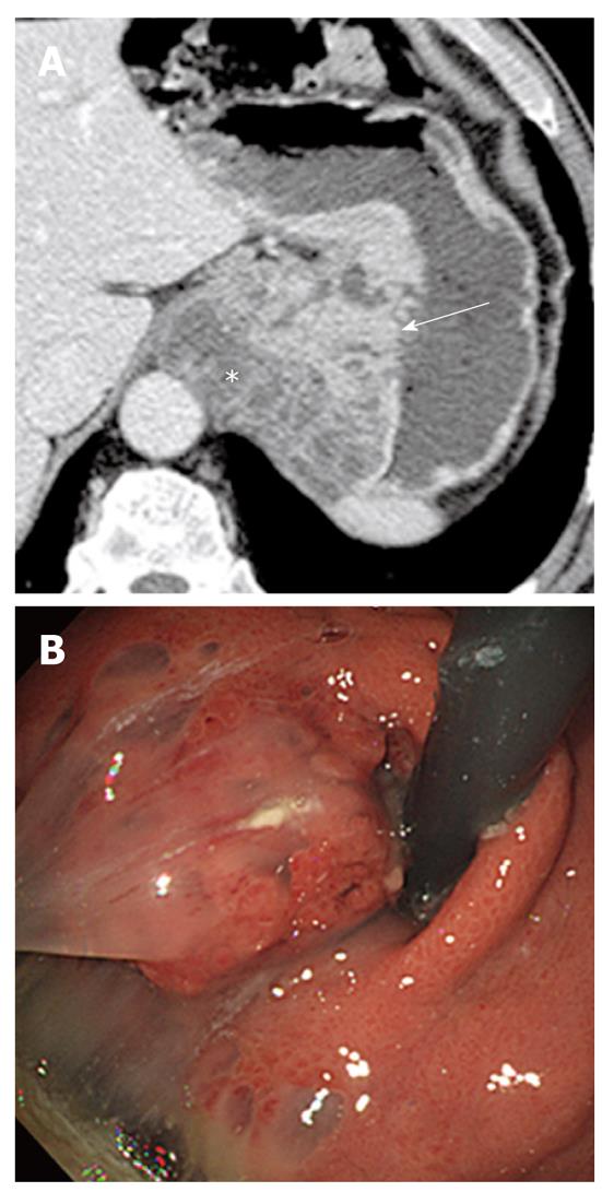
Figure 13 Mucinous carcinoma of the stomach in a 62-year-old woman.
A: Contrast-enhanced CT image shows a large mass lesion in the gastric cardia containing abundant mucin pools (asterisk) and enhanced solid portions (arrow); B: Endoscopy image shows jellylike mucin leaking from the gastric mass. CT: Computed tomography.
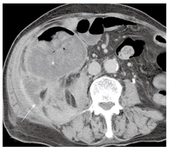
Figure 14 Mucinous adenocarcinoma of the cecum in a 69-year-old man.
Contrast-enhanced CT image shows a huge eccentric hypoattenuating mass with poor enhancement of the solid portion of the cecum (asterisk). The mass has invaded the retroperitoneum (arrow). CT: Computed tomography.
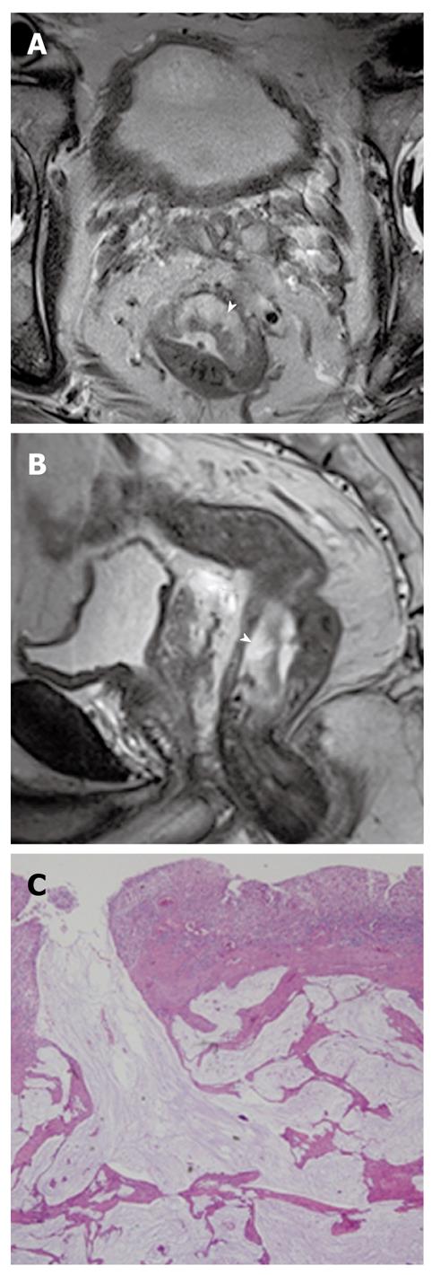
Figure 15 Mucinous adenocarcinoma of the rectum in a 63-year-old man.
A, B: Axial (A) and coronal (B) T2-weighted MR images show an area of hyperintense mucin pools (arrowhead) in the rectal mass; C: Photomicrograph image (original magnification, × 40; HE stain) shows large pools of extracellular mucin and tumor cells. MR: Magnetic resonance; HE: Hematoxylin and eosin.
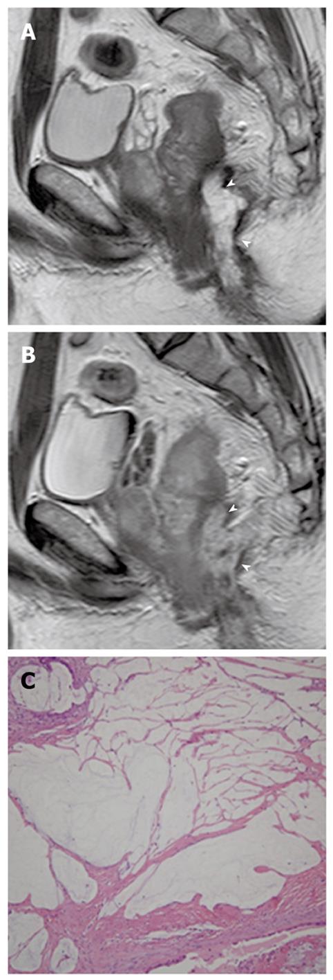
Figure 16 Perianal mucinous adenocarcinoma in a 41-year-old man.
A: Sagittal T2-weighted MR image shows a hyperintense mass (arrowheads) in the perianal area; B: Sagittal contrast-enhanced T1-weighted MR image shows mesh-like internal enhancement (arrowheads) within the mass; C: Photomicrograph image (original magnification, × 100; HE stain) shows large pools of extracellular mucin and tumor cells. MR: Magnetic resonance; HE: Hematoxylin and eosin.
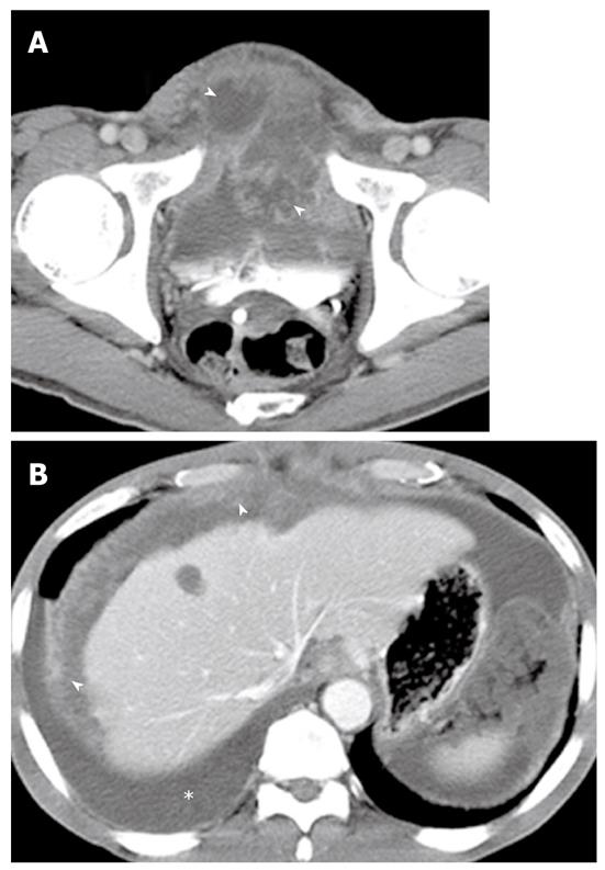
Figure 17 Intraperitoneal spread of mucinous adenocarcinoma of the urachus (also called peritoneal mucinous carcinomatosis) in a 57-year-old man.
A: Contrast-enhanced CT image shows a midline supravesical mass with heterogeneous attenuation. Within the mass are scattered low-attenuation areas (arrowheads), which represent mucin; B: Contrast-enhanced CT image of the upper abdomen shows low-attenuation mucinous ascites scalloping the liver margin. Note the right pleural effusion (asterisk) and diffuse nodular thickening of the peritoneum (arrowheads). CT: Computed tomography; MR: Magnetic resonance.
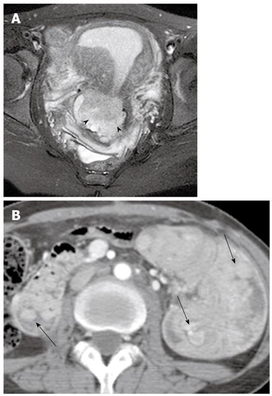
Figure 18 Adenoma malignum of the cervix in a 30-year-old woman with Peutz-Jeghers syndrome.
A: Axial T2-weighted MR image shows a multicystic lesion with a solid component (arrowheads) in the uterine cervix; B: Contrast-enhanced CT image shows multiple polyps (arrows) in the duodenum and small bowel causing intussusceptions. CT: Computed tomography; MR: Magnetic resonance.
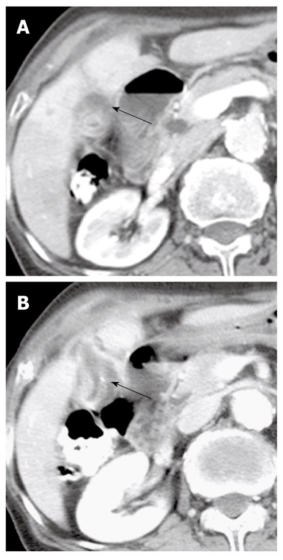
Figure 19 Mucinous carcinoma of the gallbladder in a 77-year-old woman.
A: Contrast-enhanced CT image shows localized wall thickening (arrow) in the body of the gallbladder containing a suspicious mucin pool; B: Contrast-enhanced CT image inferior to (A) shows a punctate calcification and localized wall thickening (arrow) in a mildly distended gallbladder. CT: Computed tomography.
- Citation: Lee NK, Kim S, Kim HS, Jeon TY, Kim GH, Kim DU, Park DY, Kim TU, Kang DH. Spectrum of mucin-producing neoplastic conditions of the abdomen and pelvis: Cross-sectional imaging evaluation. World J Gastroenterol 2011; 17(43): 4757-4771
- URL: https://www.wjgnet.com/1007-9327/full/v17/i43/4757.htm
- DOI: https://dx.doi.org/10.3748/wjg.v17.i43.4757


