Copyright
©2011 Baishideng Publishing Group Co.
World J Gastroenterol. Jan 28, 2011; 17(4): 526-533
Published online Jan 28, 2011. doi: 10.3748/wjg.v17.i4.526
Published online Jan 28, 2011. doi: 10.3748/wjg.v17.i4.526
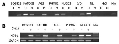
Figure 1 Silence of high in normal-1 gene expression due to methylation of high in normal-1 gene promoter in gastric carcinoma cell lines.
A: Methylation-specific polymerase chain reaction analysis of high in normal-1 (HIN-1) gene promoter methylation in five gastric carcinoma cell lines. U: Unmethylated alleles; M: Methylated alleles. In vitro methylated DNA (IVD) and DNA from normal human peripheral lymphocytes were used as methylated and unmethylated controls; B: Gastric cancer cell lines were treated with or without 5-aza-CdR (-AZ) for up to 96 h. HIN-1 mRNA levels were measured by semi-quantitative reverse transcription polymerase chain reaction analysis, and glyceraldehyde-3-phosphate dehydrogenase (GAPDH) served as control. The 1-kb marker indicated an appropriate size for the amplified products. HIN-1 expression varied among cell lines. The presence of methylation of HIN-1 corresponds directly to the loss of expression of the genes in each cell line.
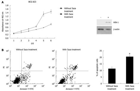
Figure 2 5-aza-2’-deoxycytidine inhibition of gastric cancer cell BCG823 viability through induction of high in normal-1 expression.
A: Gastric cancer BCG823 cells were treated with or without 5-Aza-CdR (AZ) for up to 6 d and then subjected to 3-(4,5-dimethylthiazol-2yl)-5-(3-carboxymethoxyphenyl)-2-(4-sulfophenyl)-2H-tetrazolium analysis of cell viability before and after 5-Aza-CdR treatment. The high in normal-1 (HIN-1) protein levels were measured by immunoblotting. -: Without 5aza treatment; +: With 5aza treatment; B: BCG-823 cells were subjected to FACs for apoptosis analysis. 5-aza-2’-deoxycytidine treatment inhibits BCG-823 cell proliferation (A) and induces them to undergo apoptosis (B) vs the controls (aP < 0.05).
Figure 3 Methylation-specific polymerase chain reaction analysis of high in normal-1 gene promoter methylation in gastric cancer, adjacent non-tumor tissues, and normal gastric mucosa.
A: Representative data of MS-PCR analysis of high in normal-1 (HIN-1) genes in tumor tissues (T), paired adjacent non-tumor tissues (NT) and normal gastric mucosa(N). U: Unmethylated alleles; M: Methylated alleles. In vitro methylated DNA and DNA from normal human peripheral lymphocytes were used as methylated and unmethylated controls; B: Comparison of HIN-1 gene methylation among gastric cancer (T) , adjacent non-tumor tissue (NT) and normal gastric mucosa (N). aStudent’s t test by SPSS 13.0 software, NT vs N, P = 0.005; bT vs N, P = 0.002.
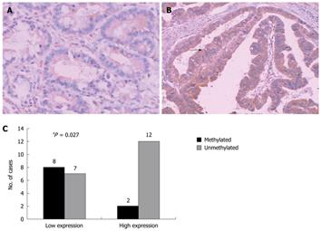
Figure 4 Immunohistochemical analysis of high in normal-1 protein expression in gastric cancer tissue samples.
A: Tumor cells with methylated alleles of high in normal-1 (HIN-1) gene promoter exhibited negative staining; B: Cancer cells without HIN-1 gene promoter methylation exhibited positive staining. HIN-1 expression in gastric cancer (membrane staining, arrow); C: The association of HIN-1 methylation with HIN-1 expression level was analyzed in 29 gastric cancers. High expression: +-+++ staining intensity with 10% or more cancer cells positively stained, otherwise it is considered as low expression. The staining intensity and percentage of staining were compared with a non-cancerous area of the same section. aPearson χ2 test or Pearson χ2 test with continuity correction by SPSS 13.0 software. A, B: IHC, × 200.
- Citation: Gong Y, Guo MZ, Ye ZJ, Zhang XL, Zhao YL, Yang YS. Silence of HIN-1 expression through methylation of its gene promoter in gastric cancer. World J Gastroenterol 2011; 17(4): 526-533
- URL: https://www.wjgnet.com/1007-9327/full/v17/i4/526.htm
- DOI: https://dx.doi.org/10.3748/wjg.v17.i4.526


