Copyright
©2011 Baishideng Publishing Group Co.
World J Gastroenterol. Oct 21, 2011; 17(39): 4365-4371
Published online Oct 21, 2011. doi: 10.3748/wjg.v17.i39.4365
Published online Oct 21, 2011. doi: 10.3748/wjg.v17.i39.4365
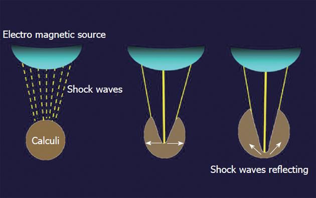
Figure 1 Principle of extracorporeal shockwave lithotripsy.
Shockwaves from the source are targeted on the calculi and these induce fragmentation.
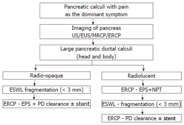
Figure 2 Protocol followed at Asian Institute of Gastroenterology, for extracorporeal shockwave lithotripsy of large pancreatic duct calculi[7].
EPS: Endoscopic pancreatic sphincterotomy; US: Ultrasound; EUS: Endoscopic ultrasound; MRCP: Magnetic resonance cholangiopancreatography; ERCP: Endoscopic retrograde cholangiopancreatography; PD: Pancreatic duct; ESWL: Extracorporeal shock wave lithotripsy; NPT: Naso-pancreatic tube.
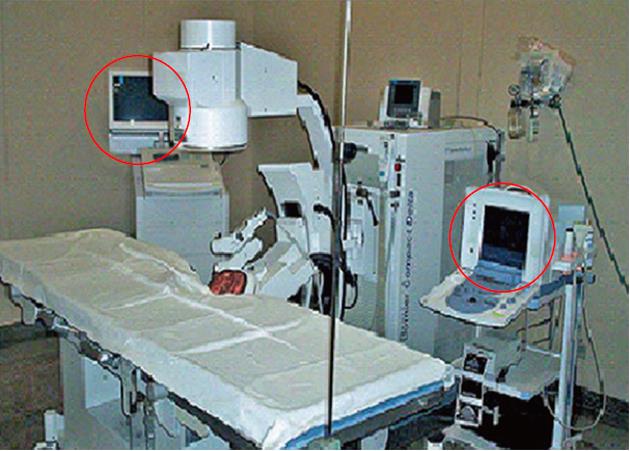
Figure 3 Third-generation lithotripter with fluoroscopic and ultrasound imaging facility.
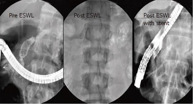
Figure 4 Large pancreatic calculi in head and genu, cleared by extracorporeal shockwave lithotripsy followed by pancreatic stenting.
ESWL: Extracorporeal shockwave lithotripsy.
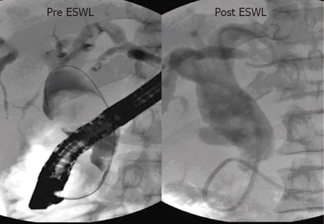
Figure 5 Large pancreatic calculi in head.
Post extracorporeal shockwave lithotripsy (ESWL) reduction in diameter of main pancreatic duct.
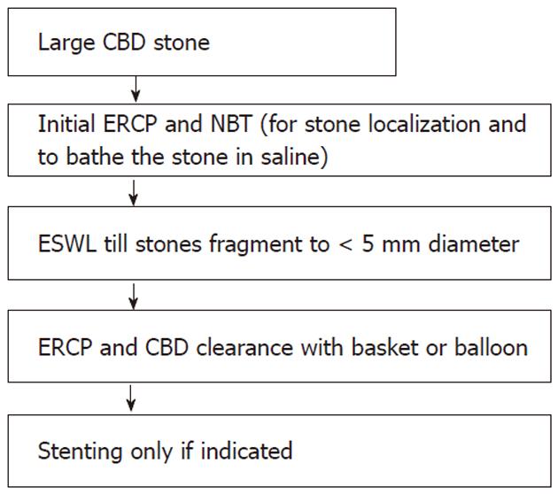
Figure 6 Protocol for extracorporeal shockwave lithotripsy of large common bile duct calculi.
CBD: Common bile duct; ERCP: Endoscopic retrograde cholangiopancreatography; NBT: Nasobiliary tube; ESWL: Extracorporeal shockwave lithotripsy.
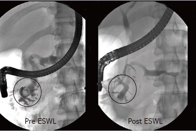
Figure 7 Large common bile duct calculi with narrow distal common bile duct.
Good fragmentation achieved with extracorporeal shockwave lithotripsy (ESWL).
- Citation: Tandan M, Reddy DN. Extracorporeal shock wave lithotripsy for pancreatic and large common bile duct stones. World J Gastroenterol 2011; 17(39): 4365-4371
- URL: https://www.wjgnet.com/1007-9327/full/v17/i39/4365.htm
- DOI: https://dx.doi.org/10.3748/wjg.v17.i39.4365


