©2011 Baishideng Publishing Group Co.
World J Gastroenterol. Sep 28, 2011; 17(36): 4118-4123
Published online Sep 28, 2011. doi: 10.3748/wjg.v17.i36.4118
Published online Sep 28, 2011. doi: 10.3748/wjg.v17.i36.4118
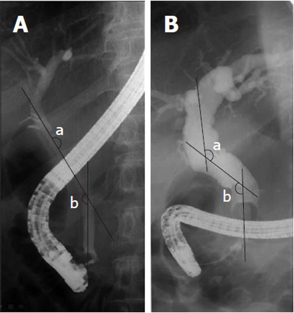
Figure 1 Cholangiographic configuration of (A) usual extrahepatic bile duct and (B) deformed bile duct.
The imaginary line was drawn along the center of the bile duct and each internal angle was measured at the angulation of the proximal (a) and distal (b) bile duct level respectively. The values of both angles were added together (a + b).
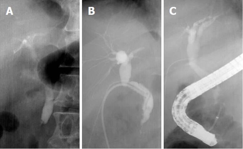
Figure 2 The bile duct angulation caused by T-tube choledochostomy.
A: Extrahepatic bile duct configuration before T-tube choledochostomy; B: Bile duct configuration during T-tube drainage; C: Bile duct configuration altered after T-tube drainage. This change of cholangiographic findings showed that T-tube drainage would have induced bile duct angulation.
- Citation: Seo DB, Bang BW, Jeong S, Lee DH, Park SG, Jeon YS, Lee JI, Lee JW. Does the bile duct angulation affect recurrence of choledocholithiasis? World J Gastroenterol 2011; 17(36): 4118-4123
- URL: https://www.wjgnet.com/1007-9327/full/v17/i36/4118.htm
- DOI: https://dx.doi.org/10.3748/wjg.v17.i36.4118


