©2011 Baishideng Publishing Group Co.
World J Gastroenterol. Sep 21, 2011; 17(35): 3986-3993
Published online Sep 21, 2011. doi: 10.3748/wjg.v17.i35.3986
Published online Sep 21, 2011. doi: 10.3748/wjg.v17.i35.3986
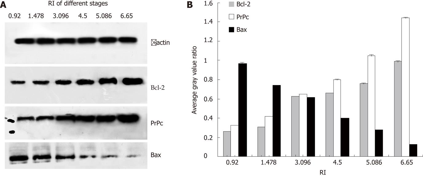
Figure 1 Expressions of PrPc, Bcl-2 and Bax in gastric cancer cells resistant to different concentrations of adriamycin.
Analysis indicated that the expressions of PrPc and Bcl-2 were markedly increased (P < 0.01), but that of Bax was significantly reduced (P < 0.01), which were in an adriamycin concentration dependent manner. Resistance index was 3.096 when the adriamycin concentration was 0.18 μg/mL. PrPc: Prion protein; RI: Resistence index.
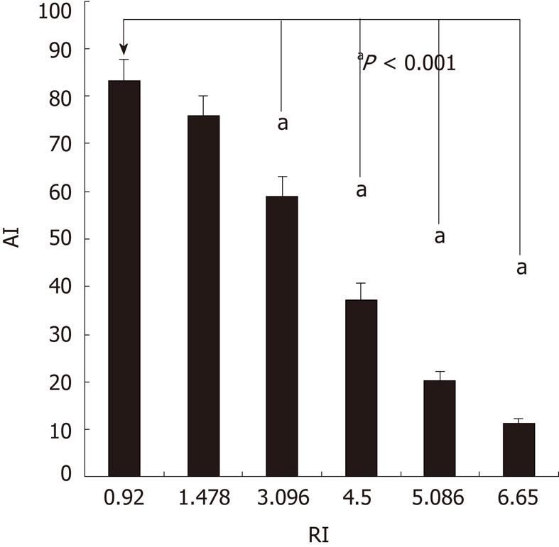
Figure 2 Apoptosis index of gastric cancer cells resistant to adriamycin of different concentrations.
In the SGC7901 cells resistant to 0.18 μg/mL adriamycin, the anti-apoptotic capability was significantly increased with the resistence index of 3.096 (aP < 0.001) when compared with SGC7901 cells without adriamycin treatment. AI: Apoptosis index; RI: Resistence index.
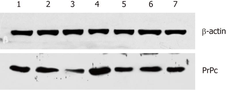
Figure 3 Prion protein expression in different gastric cancer cell lines.
1: KATOIII; 2: BGC-823; 3: GES; 4: HGC-27; 5: AGS; 6: SGC7901; 7: MGC80. Results showed that the highest prion protein (PrPc) expression was found in undifferentiated gastric cancer cells (HGC-27), followed by poorly differentiated gastric cancer cells (KATOIII), and the lowest PrPc expression in human gastric epithelial immortalized GES-1 cells. PrPc: Prion protein.
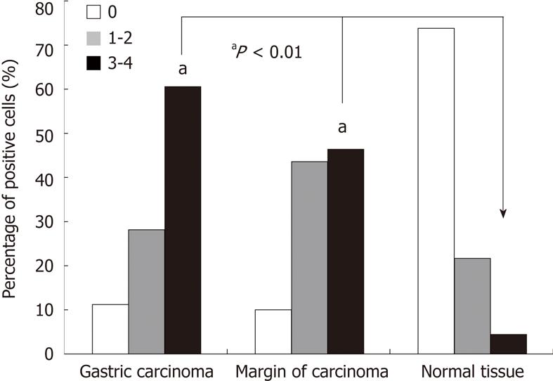
Figure 4 Prion protein expression in gastric cancers, precancerous lesions and normal tissues.
χ2 test revealed that the number of prion protein positive cells in gastric cancers and precancerous lesions were dramatically increased when compared with normal tissues (aP < 0.01). PrPc: Prion protein.
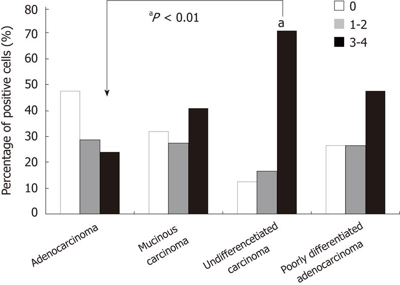
Figure 5 Prion protein expression in different type of gastric cancer.
The proportion of prion protein positive cells in undifferentiated cancer was significantly higher than that in adenocarcinoma (aP < 0.01), but no significant difference in PrPc positive cells was found between mucinous carcinoma and poorly differentiated adenocarcinoma. PrPc: Prion protein.
Figure 6 Prion protein expression in gastric cancers and normal tissues.
The prion protein (PrPc) expression in gastric adenocarcinoma and mixed carcinoma was relatively low, and no significant difference in PrPc expression was noted between the remaining types of gastric cancer. Furthermore, PrPc expression in normal gastric tissues was much lower than that in gastric cancers. PrPc: Prion protein. 1: Tissue of atrophic gastritis; 2: Tissue of undifferentiated gastric cancer; 3: Tissue of gastric adenocarcinoma; 4: Tissue of mixed type carcinoma; 5: Tissue of poorly differentiated gastric cancer; 6-10: Corresponding normal gastric tissue.
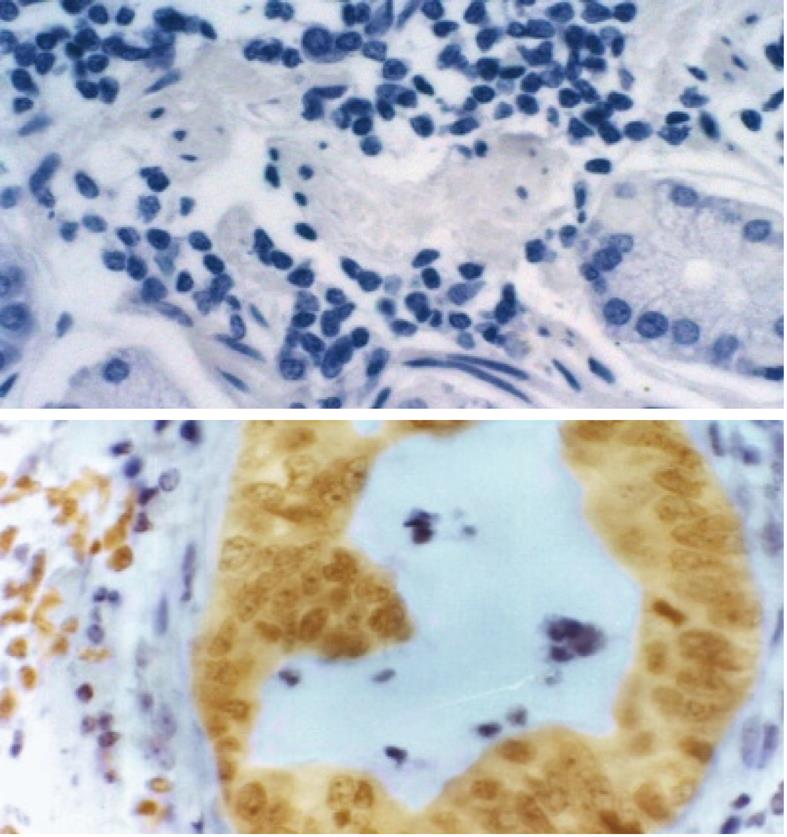
Figure 7 Prion protein expression in human gastric cancer (× 200).
A: Negative control; B: Positive results.
- Citation: Wang JH, Du JP, Zhang YH, Zhao XJ, Fan RY, Wang ZH, Wu ZT, Han Y. Dynamic changes and surveillance function of prion protein expression in gastric cancer drug resistance. World J Gastroenterol 2011; 17(35): 3986-3993
- URL: https://www.wjgnet.com/1007-9327/full/v17/i35/3986.htm
- DOI: https://dx.doi.org/10.3748/wjg.v17.i35.3986


