Copyright
©2011 Baishideng Publishing Group Co.
World J Gastroenterol. Sep 14, 2011; 17(34): 3948-3952
Published online Sep 14, 2011. doi: 10.3748/wjg.v17.i34.3948
Published online Sep 14, 2011. doi: 10.3748/wjg.v17.i34.3948
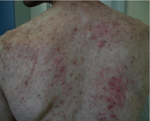
Figure 1 Maculopapular rash (“urticaria pigmentosa”) in a systemic mastocytosis patient.
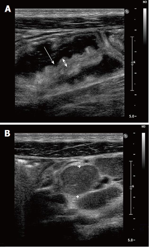
Figure 2 Abdominal ultrasound.
A: Small bowel dilatation and wall edema at ultrasonography (US). B: Abdominal lymphadenopathy at US (crosses refer to lymph node enlargement, 5 cm).
Figure 3 Colon biopsy in a systemic mastocytosis patient.
A: Diffuse mast cell (MC) infiltrate (Hematoxylin-eosin, × 10); B: The dense infiltrate is represented by MCs, whose detection is increased by positive immunohistochemical marker CD117; C: The dense infiltrate is represented by MCs, whose detection is increased by positive immunohistochemical marker CD25.
Figure 4 Bone marrow biopsy in a systemic mastocytosis patient.
A: Diffuse mast cell (MC) infiltrate (Hematoxylin-eosin, × 10); B: The dense infiltrate is represented by MCs, whose detection is increased by positive immunohistochemical marker CD117; C: The dense infiltrate is represented by MCs, whose detection is increased by positive immunohistochemical marker CD25.
- Citation: Elvevi A, Grifoni F, Branchi F, Gianelli U, Conte D. Severe chronic diarrhea and maculopapular rash: A case report. World J Gastroenterol 2011; 17(34): 3948-3952
- URL: https://www.wjgnet.com/1007-9327/full/v17/i34/3948.htm
- DOI: https://dx.doi.org/10.3748/wjg.v17.i34.3948


