©2011 Baishideng Publishing Group Co.
World J Gastroenterol. Aug 21, 2011; 17(31): 3623-3629
Published online Aug 21, 2011. doi: 10.3748/wjg.v17.i31.3623
Published online Aug 21, 2011. doi: 10.3748/wjg.v17.i31.3623
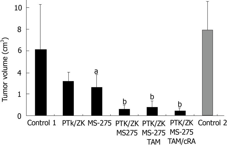
Figure 1 Macroscopic tumor growth.
The results are given as absolute values. aP = 0.025, MS-275 vs control 1; bP = 0.005, ZK/PTK/MS vs control 1. PTK/ZK: PTK787/ZK222584 (Vatalanib®); TAM: Tamoxifen; cRA: 9-cis-retinoic acid.
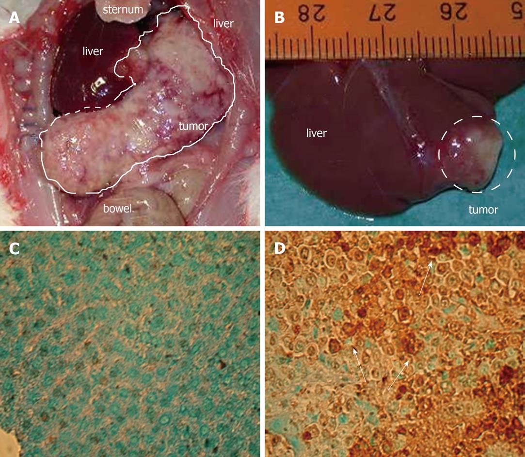
Figure 2 Macroscopic tumor growth and TdT-mediated dUTP-biotin nick end labeling assay.
A: Control 1, large tumor volume. B: PTK787/ZK222584 (PTK/ZK) + MS-275, small tumor volume. C: Control 1, (mediated dUTP-biotin nick end labeling) TUNEL assay for apoptotic cells (dark brown). D: PTK/ZK + MS-275: TUNEL assay for apoptotic cells (dark brown).
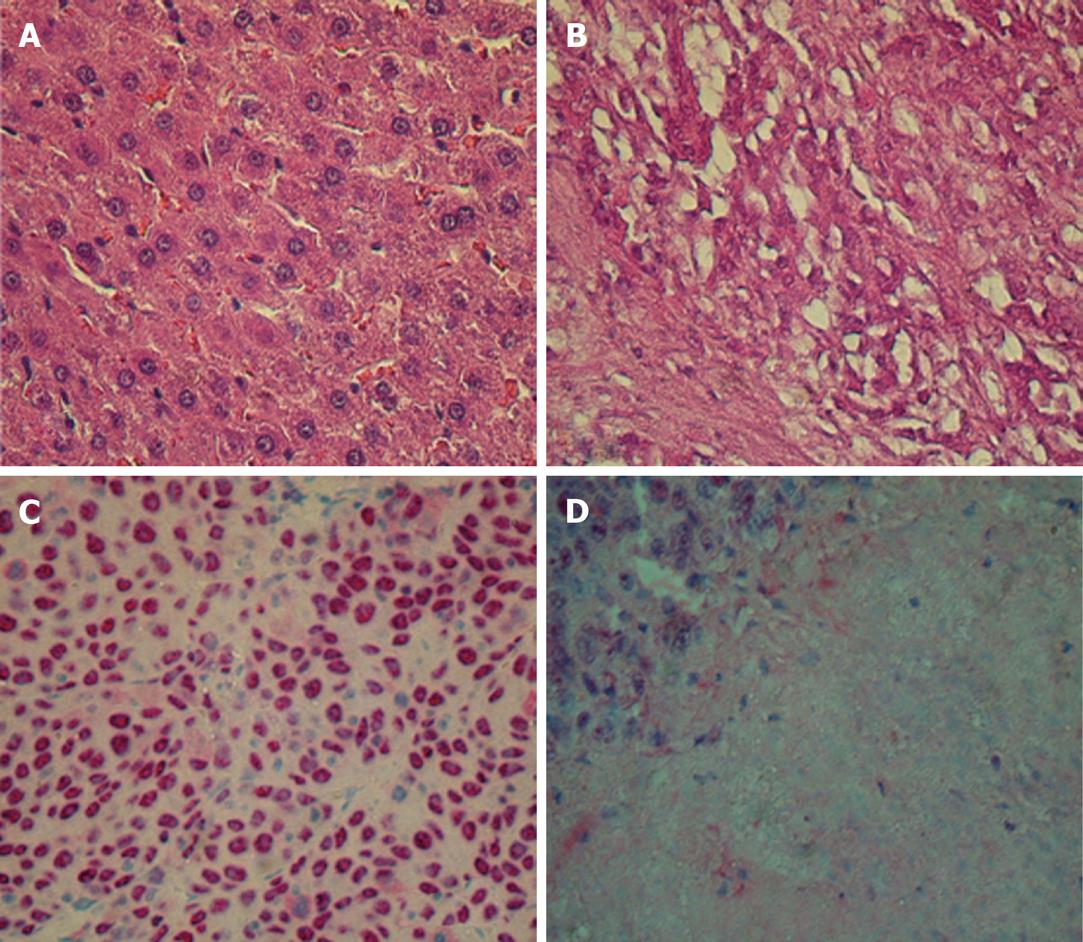
Figure 3 Hematoxylin-eosin and proliferating cell nuclear antigen staining.
A: Control 1, hematoxylin-eosin staining. B: Quadruple therapy, disintegrating cells, necrosis. C: Control 1, proliferating cell nuclear antigen (PCNA) staining for proliferating cells. D: Quadruple therapy, PCNA staining, reduced number of cells, and large necrotic area.
- Citation: Ganslmayer M, Zimmermann A, Zopf S, Herold C. Combined inhibitors of angiogenesis and histone deacetylase: Efficacy in rat hepatoma. World J Gastroenterol 2011; 17(31): 3623-3629
- URL: https://www.wjgnet.com/1007-9327/full/v17/i31/3623.htm
- DOI: https://dx.doi.org/10.3748/wjg.v17.i31.3623


