Copyright
©2011 Baishideng Publishing Group Co.
World J Gastroenterol. Aug 14, 2011; 17(30): 3544-3553
Published online Aug 14, 2011. doi: 10.3748/wjg.v17.i30.3544
Published online Aug 14, 2011. doi: 10.3748/wjg.v17.i30.3544
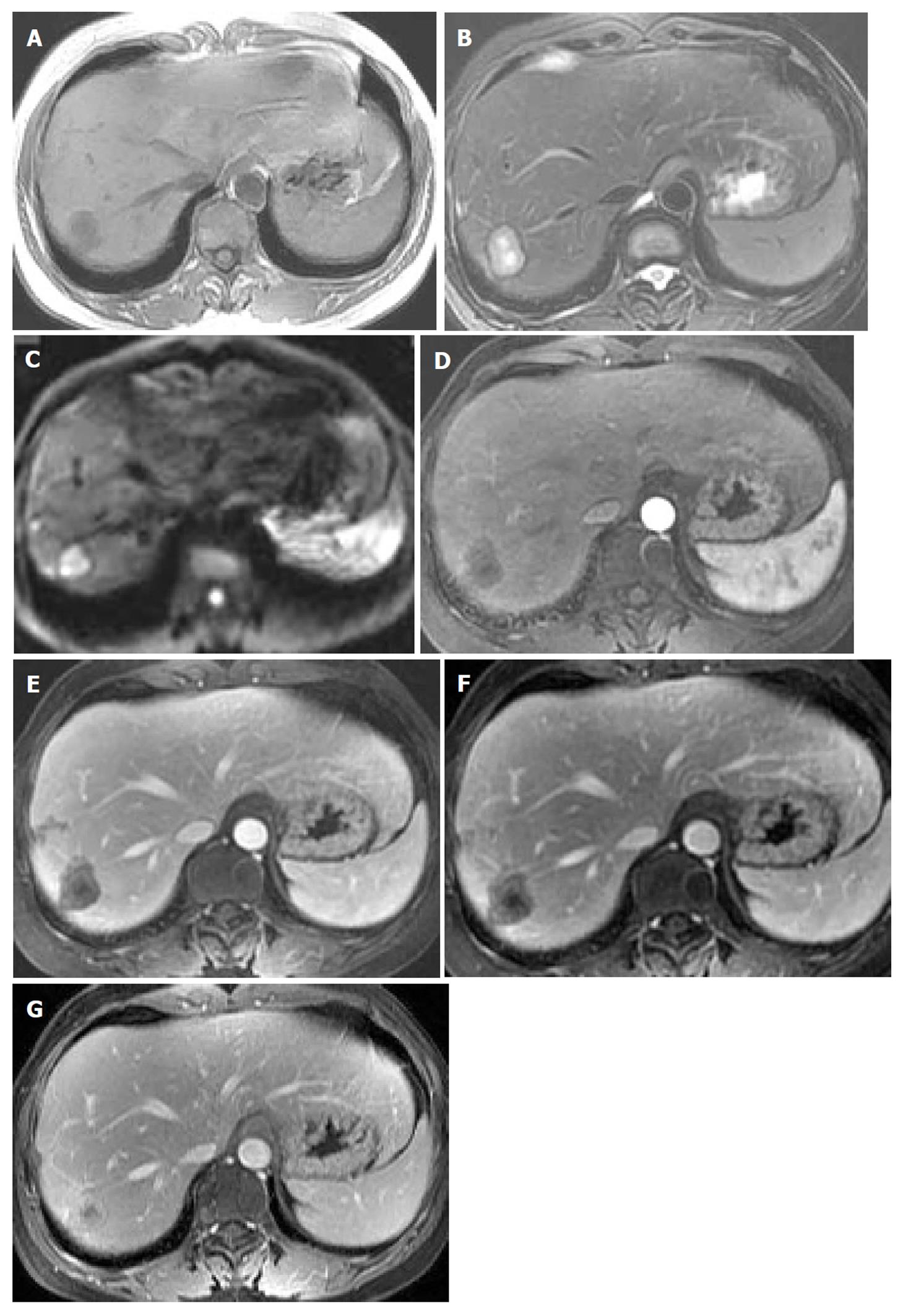
Figure 1 Multifocal hepatic epithelioid hemangioendothelioma in a 27-year-old male.
(A) Unenhanced axial computed tomography scan of liver shows multiple discrete masses, which are round with a low density and peripheral iso-density. (B-C) Partial lesions show peripheral ring-like enhancement in the arterial phase (B) with even stronger enhancement in the portal venous phase (C).
Figure 2 Multifocal hepatic epithelioid hemangioendothelioma in a 48-year-old female.
Precontrast axial magnetic resonance imaging scan of the liver shows multiple lesions of low signal intensity with peripheral faint hyperintensity on T1WI (A), high signal intensity with peripheral hypointensity and an area of evident hyperintensity in the center of one lesion on T2WI (B), and hyperintensity with peripheral hypointensity on diffusion weighted imaging (C). Lesions show peripheral ring-like enhancement in the arterial phase (D), and heterogeneously progressive reinforcement in the portal venous phase (E), equilibrium phase (F) and it approaches isointensity to liver parenchyma in the delayed phase (G). There is an association with an area of unenhanced necrosis in the center, which looks like a conspicuous “target” with an inner low intensity, in-between high intensity and outer lower intensity layers.
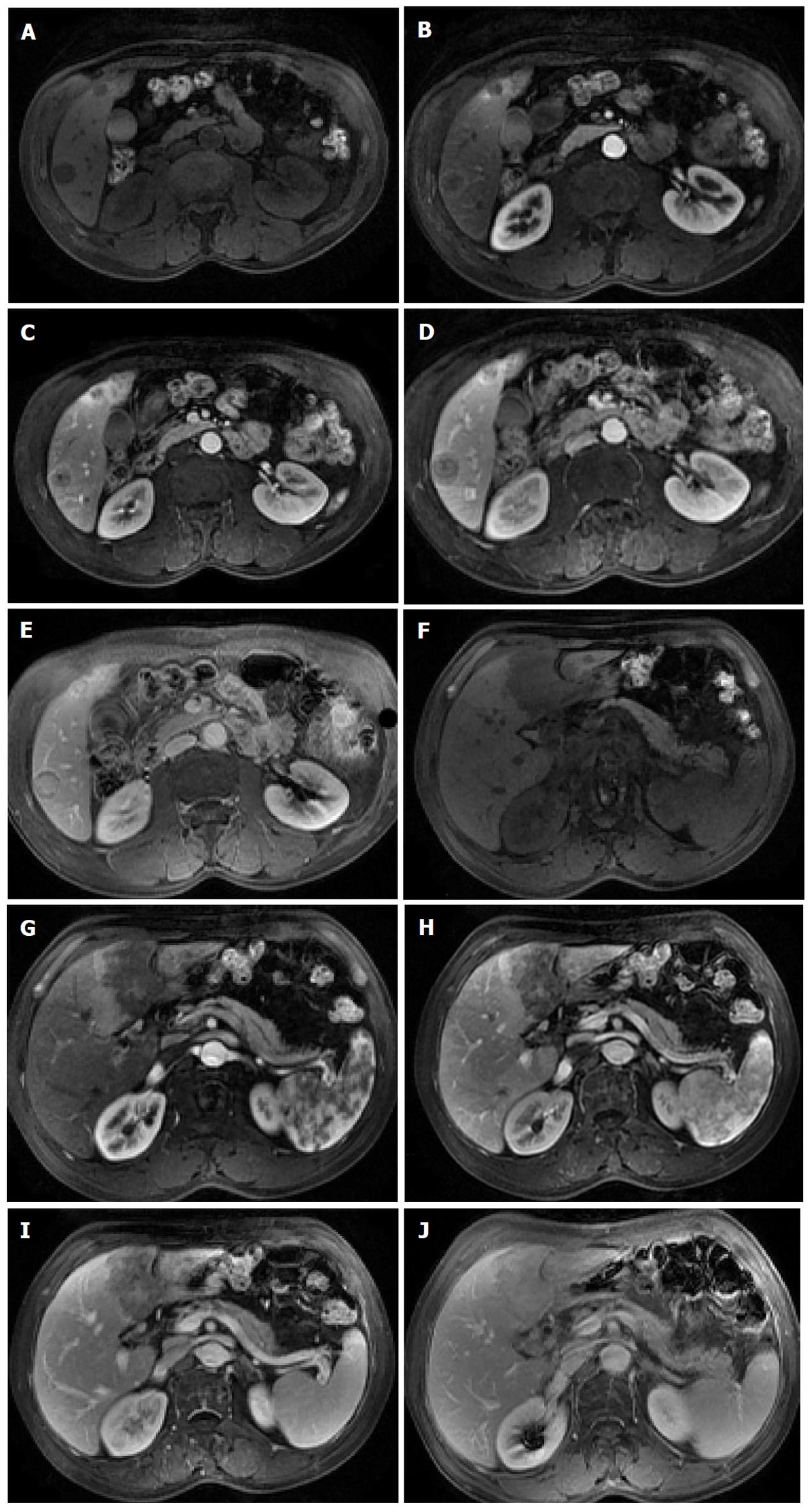
Figure 3 Multifocal hepatic epithelioid hemangioendothelioma in a 60-year-old man.
Contrast-enhanced multiple-phase magnetic resonance imaging shows a progressive-enhancement target sign in the lesions in segment VI of the liver (A-E) and lamellar lesions with progressive enhancement in the left lobe of the liver (F-J).
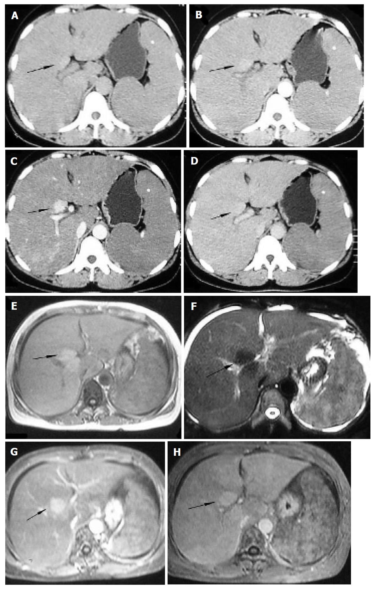
Figure 4 Diffuse hepatic epithelioid hemangioendothelioma in a 48-year-old woman.
Plain computed tomography and magnetic resonance imaging manifests an obviously enlarged liver with a large nodule (black arrow) appearing slightly hyperdense relative to normal liver parenchyma (A), isointense on T1WI (E) and hypointense on T2WI (F), with slight enhancement in the arterial phase (B), evident enhancement in the portal venous phase (C and G) and iso-density/intensity in the equilibrium phase (D and H), associated with splenomegaly and ascites (F).
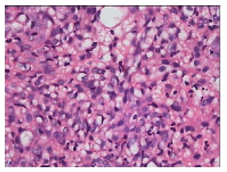
Figure 5 Photomicrography shows epithelioid cells with intracytoplasmic lumina, occasionally containing red blood cells, appearing as signet ring–like structures (hematoxylin and eosin, HE × 200).
Figure 6 Immunohistochemical staining shows that the tumor cells are positive for factor VIII-related antigen (A), CD34 (B) and CD31 (C) (× 250).
- Citation: Chen Y, Yu RS, Qiu LL, Jiang DY, Tan YB, Fu YB. Contrast-enhanced multiple-phase imaging features in hepatic epithelioid hemangioendothelioma. World J Gastroenterol 2011; 17(30): 3544-3553
- URL: https://www.wjgnet.com/1007-9327/full/v17/i30/3544.htm
- DOI: https://dx.doi.org/10.3748/wjg.v17.i30.3544


