©2011 Baishideng Publishing Group Co.
World J Gastroenterol. Jul 7, 2011; 17(25): 3066-3068
Published online Jul 7, 2011. doi: 10.3748/wjg.v17.i25.3066
Published online Jul 7, 2011. doi: 10.3748/wjg.v17.i25.3066
Figure 1 Gastric carcinoid.
A: Infiltration of the muscularis mucosae; Hematoxylin-eosin stain. Magnification, × 50; B: Tumor and normal mucosa adjacent to tumor immunostained for VMAT-2. Virtually all tumor cells positive. Magnification, × 100; C: Tumor immunostained for Ki-67. < 1% tumor cells positive. Magnification, × 200.
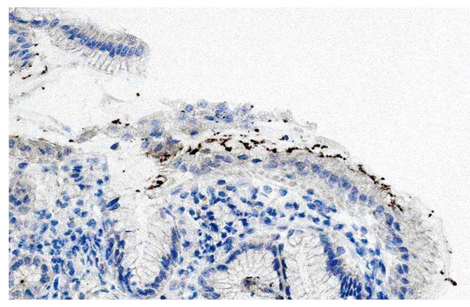
Figure 2 Signs of Helicobacter pylori infection in biopsy from antral mucosa.
Magnification, × 200.
-
Citation: Antonodimitrakis P, Tsolakis A, Welin S, Kozlovacki G, Öberg K, Granberg D. Gastric carcinoid in a patient infected with
Helicobacter pylori : A new entity? World J Gastroenterol 2011; 17(25): 3066-3068 - URL: https://www.wjgnet.com/1007-9327/full/v17/i25/3066.htm
- DOI: https://dx.doi.org/10.3748/wjg.v17.i25.3066


