Copyright
©2011 Baishideng Publishing Group Co.
World J Gastroenterol. Jun 7, 2011; 17(21): 2632-2640
Published online Jun 7, 2011. doi: 10.3748/wjg.v17.i21.2632
Published online Jun 7, 2011. doi: 10.3748/wjg.v17.i21.2632
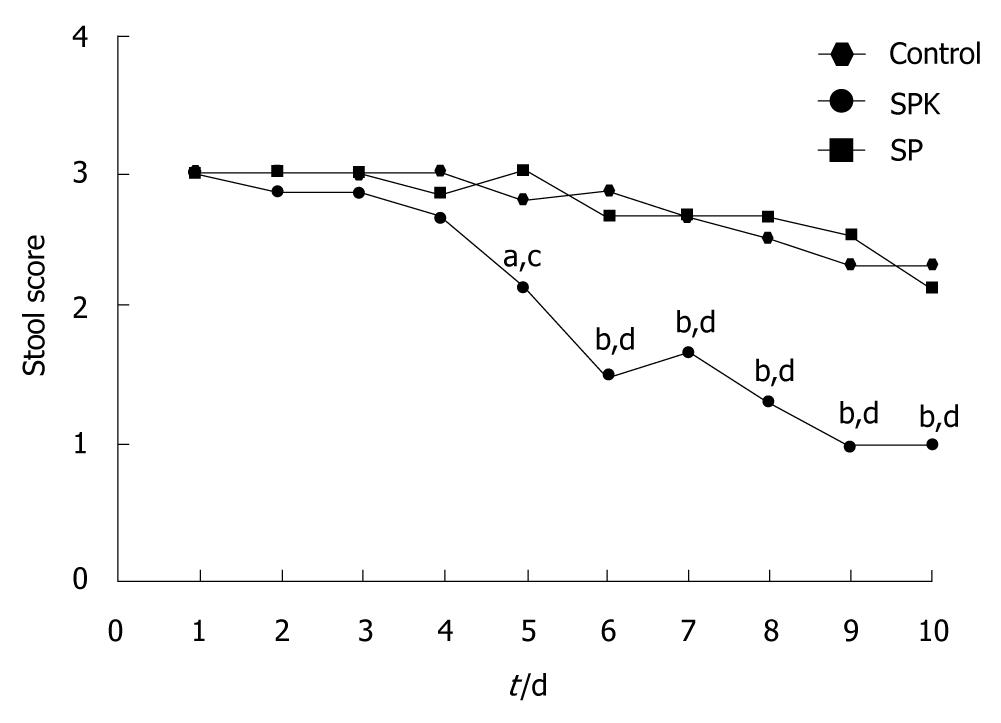
Figure 1 Stool score in each group after administration.
Stool score decreased significantly on day 5 in SPK group, and recovered to normal on day 9. Data are presented as mean ± SD, n = 6. aP < 0.05, bP < 0.01 vs SP group; cP < 0.05, dP < 0.01 vs control group at the indicated time point. SP: Attenuated Salmonella typhimurium Ty21a strain; SPK: Attenuated Salmonella typhimurium Ty21a strain carrying human KGF gene.
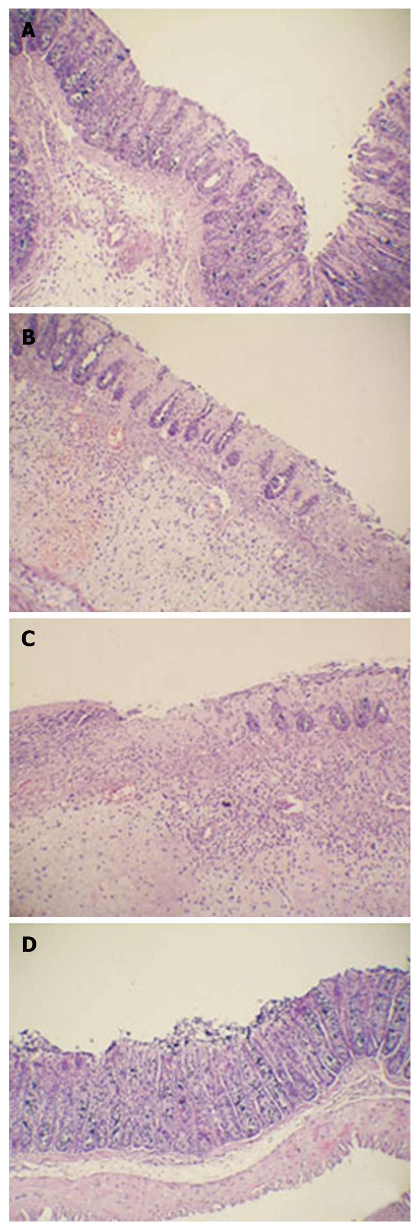
Figure 2 Histological sections of the colon on day 10 after treatment.
Inflammatory cell infiltration and edema decreased evidently in attenuated Salmonella typhimurium Ty21a strain carrying human keratinocyte growth factor gene (SPK) group compared with attenuated Salmonella typhimurium Ty21a strain (SP) and control groups. Glands arranged more regularly and there was not obvious hyperemia in the lamina propria in SPK group. A: A representative colon from SPK group; B: A representative colon from SP group; C: A representative colon from control group. D: A representative colon from normal group (HE stain, × 200).
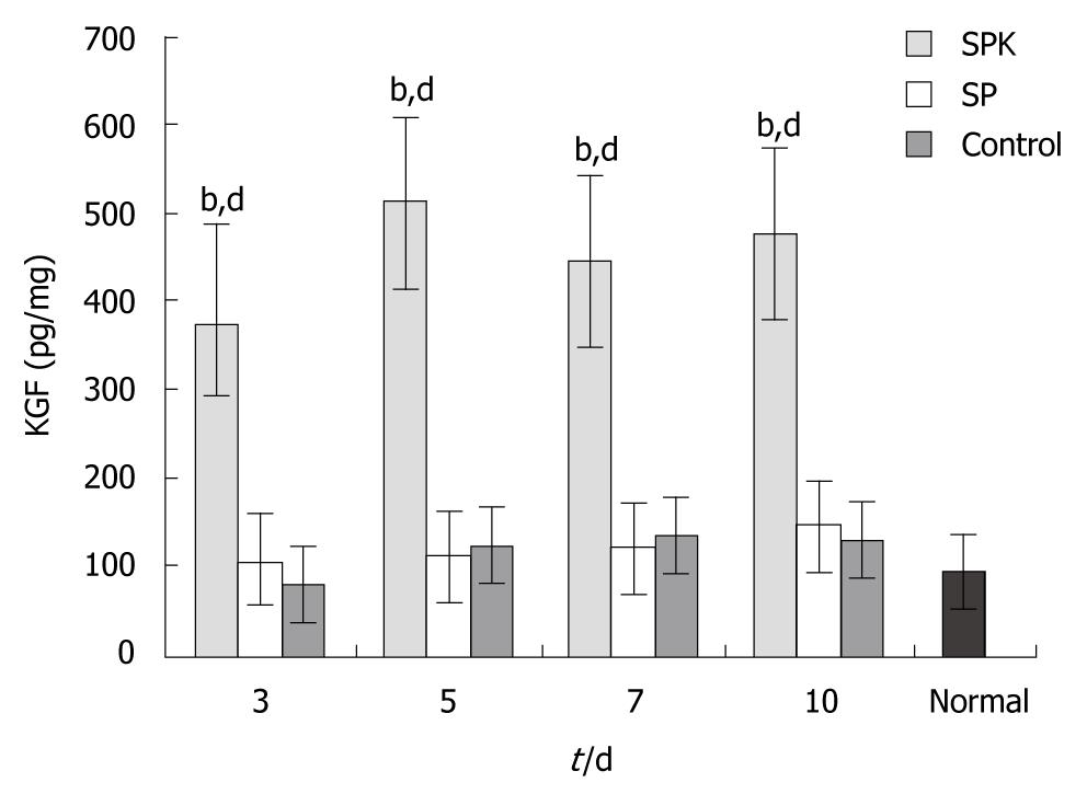
Figure 3 Keratinocyte growth factor concentrations in the homogenate of colon tissues.
The concentration of keratinocyte growth factor (KGF) in the homogenate was measured by Enzyme-linked immunosorbent assay after administration, and the expression of KGF increased obviously in attenuated Salmonella typhimurium Ty21a strain carrying human KGF gene (SPK) group. Data are presented as mean ± SD, n = 6. bP < 0.01 vs SP group at the same time point; dP < 0.01 vs control group at the same time point. SP: Attenuated Salmonella typhimurium Ty21a strain.
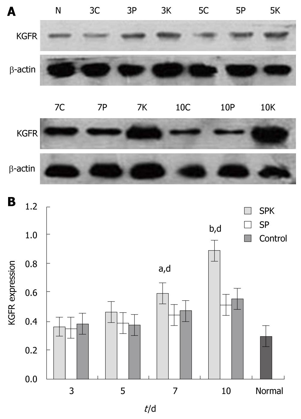
Figure 4 Western blotting of keratinocyte growth factor receptor in colon tissues.
The expression of keratinocyte growth factor receptor (KGFR) in the colon tissues was measured by Western blotting after drug administration. The expression of KGFR on days 7 and 10 increased obviously in attenuated Salmonella typhimurium Ty21a strain carrying human keratinocyte growth factor gene group (SPK) compared with attenuated Salmonella typhimurium Ty21a strain (SP) and control groups. A: Western blotting analysis of KGFR. N: normal group; 3C, 3P and 3K represents control, SP and SPK groups, respectively, on day 3 of drug administration; 5C, 5P and 5K, on day 5; 7C, 7P and 7K, on day 7; 10 C, 10P and 10K, on day 10; B: Analysis of Western blotting of KGFR. The density of the bands was quantified using image-pro plus 6.0 software, and the data are presented as mean ± SD, n = 6. aP < 0.05, bP < 0.01 vs control group; dP < 0.01, vs SP group at the same time point.
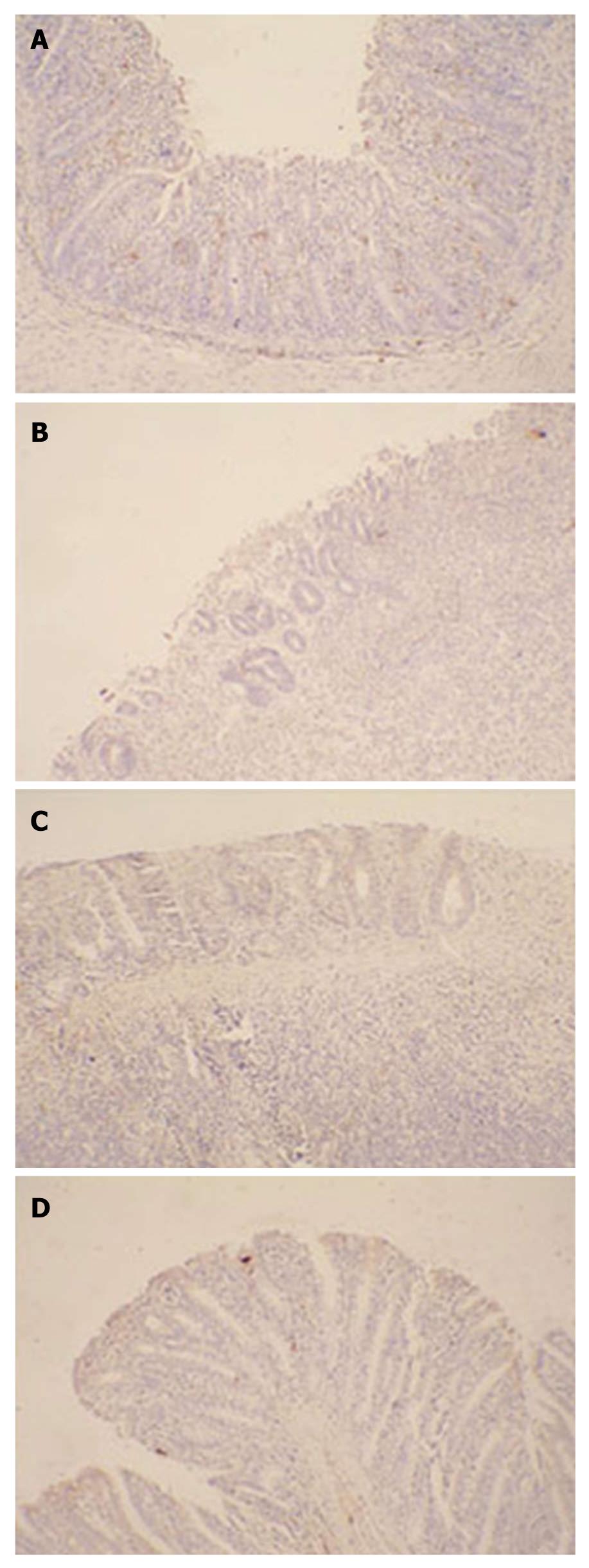
Figure 5 Immunohistochemistry of keratinocyte growth factor receptor in colon tissues.
The expression of the keratinocyte growth factor receptor (KGFR) protein was confirmed by immunohistochemistry with a rabbit anti-rat KGFR antibody as the primary antibody and a horse radish peroxidase-conjugated goat anti-rabbit antibody as the secondary antibody, and the brown color was considered to be positive staining. The expression of KGFR elevated significantly in attenuated Salmonella typhimurium Ty21a strain carrying human keratinocyte growth factor gene (SPK) group which was consistent with the results of Western blotting, and KGFR is located mainly in epithelial lamina. A: Representative wound tissue from the SPK group; B: Representative wound tissue from attenuated Salmonella typhimurium Ty21a strain group; C: Representative wound tissue from control group; D: Representative wound tissue from normal group (× 200).
Figure 6 Immunohistochemistry of Ki67 in colon tissues.
The expression of the Ki67 protein was detected by immunohistochemistry with a rabbit anti-rat Ki67 antibody as the primary antibody and a horse radish peroxidase-conjugated goat anti-rabbit antibody as the secondary antibody, and the brown color was considered to be positive staining. The expression of Ki67 in damaged colonic tissues increased evidently in the group administered with attenuated Salmonella typhimurium Ty21a strain carrying human keratinocyte growth factor gene (SPK) strain, indicating the proliferation of colonic epithelial cells. A: Representative wound tissue from SPK group; B: Representative wound tissue from attenuated Salmonella typhimurium Ty21a strain group; C: Representative wound tissue from control group; D: Representative wound tissue from normal group (× 200).
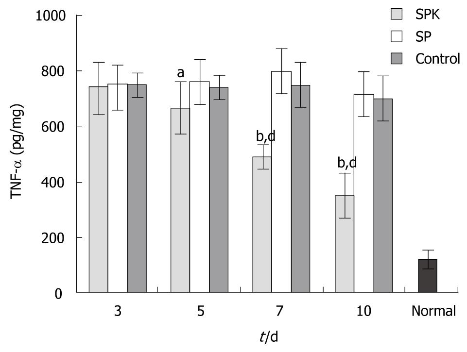
Figure 7 Tumor necrosis factor-α concentration in homogenate of colon tissues.
The concentration of tumor necrosis factor (TNF)-α in the homogenate was measured by enzyme-linked immunosorbent assay after administration. The expression of TNF-α decreased significantly in attenuated Salmonella typhimurium Ty21a strain carrying human keratinocyte growth factor gene (SPK) group from day 5 to day 10. Data are presented as mean ± SD, n = 6. aP < 0.05, bP < 0.01 vs attenuated Salmonella typhimurium Ty21a strain (SP) group; dP < 0.01 vs control group at the same time point.
- Citation: Liu CJ, Jin JD, Lv TD, Wu ZZ, Ha XQ. Keratinocyte growth factor gene therapy ameliorates ulcerative colitis in rats. World J Gastroenterol 2011; 17(21): 2632-2640
- URL: https://www.wjgnet.com/1007-9327/full/v17/i21/2632.htm
- DOI: https://dx.doi.org/10.3748/wjg.v17.i21.2632


