Copyright
©2011 Baishideng Publishing Group Co.
World J Gastroenterol. May 21, 2011; 17(19): 2417-2423
Published online May 21, 2011. doi: 10.3748/wjg.v17.i19.2417
Published online May 21, 2011. doi: 10.3748/wjg.v17.i19.2417
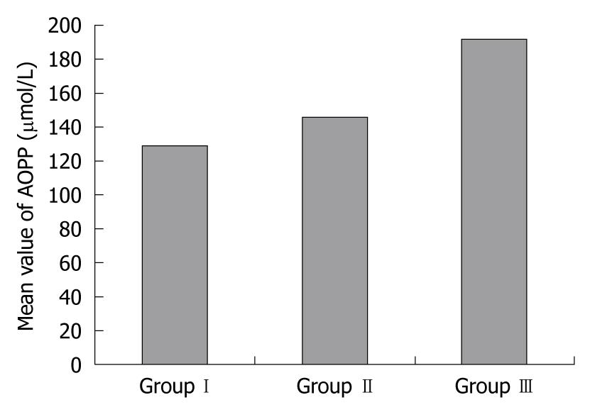
Figure 1 Comparison of advanced oxidation protein product levels in groups I, II and III.
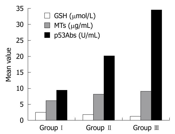
Figure 2 Comparison of glutathione, metallothioneins, and p53 antibodies levels in all studied groups.
GSH: Glutathione; MTs: Metallothioneins; p53Abs: p53 antibodies.
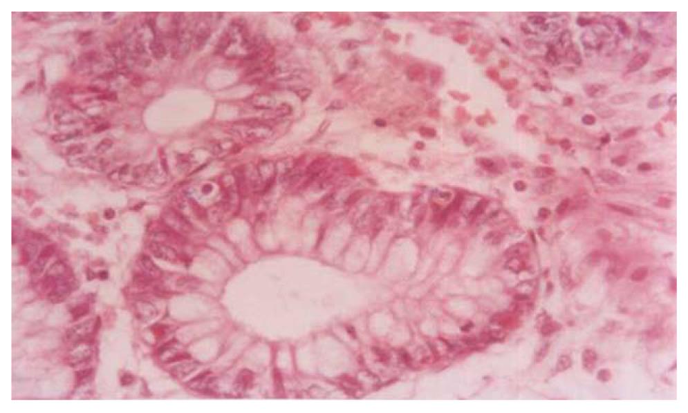
Figure 3 Photomicrograph of ulcerative colitis mucosa without dysplasia showing acute inflammatory cells (stromal) and multiple apoptotic bodies (400 ×).
Hematoxylin and eosin stain.
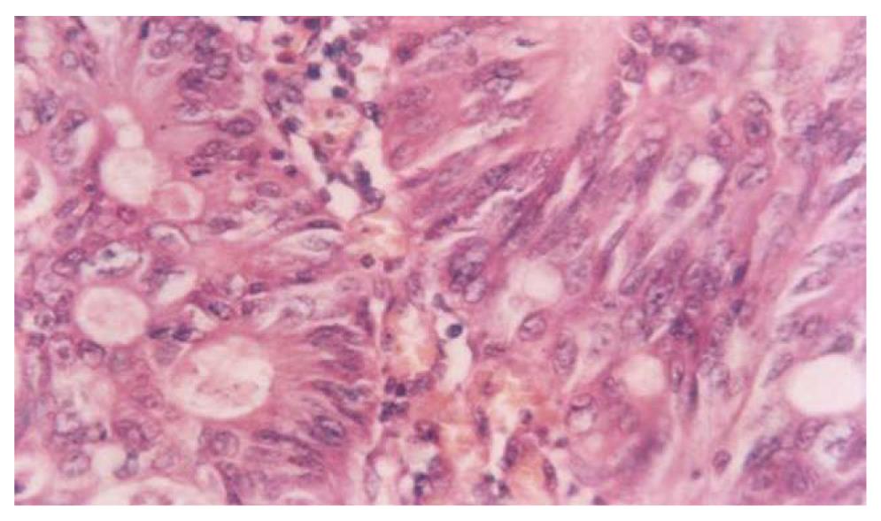
Figure 4 Photomicrograph of ulcerative colitis with dysplasia showing hyperchromatic nuclei shrunken cells, cytoplasmic organelles and inclusions (400 ×).
Hematoxylin and eosin stain.
- Citation: Hamouda HE, Zakaria SS, Ismail SA, Khedr MA, Mayah WW. p53 antibodies, metallothioneins, and oxidative stress markers in chronic ulcerative colitis with dysplasia. World J Gastroenterol 2011; 17(19): 2417-2423
- URL: https://www.wjgnet.com/1007-9327/full/v17/i19/2417.htm
- DOI: https://dx.doi.org/10.3748/wjg.v17.i19.2417


