Copyright
©2011 Baishideng Publishing Group Co.
World J Gastroenterol. Jan 7, 2011; 17(1): 49-52
Published online Jan 7, 2011. doi: 10.3748/wjg.v17.i1.49
Published online Jan 7, 2011. doi: 10.3748/wjg.v17.i1.49
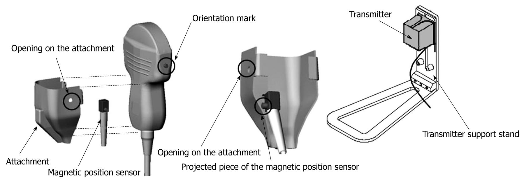
Figure 1 The probe and the magnetic sensor are assembled with careful attention to the positions of the orientation marks.
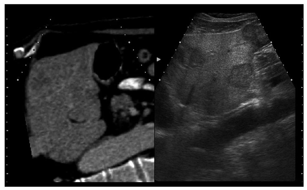
Figure 2 The left liver lobe ultrasound and computed tomography images are visualized at the same time in real-time virtual sonography.
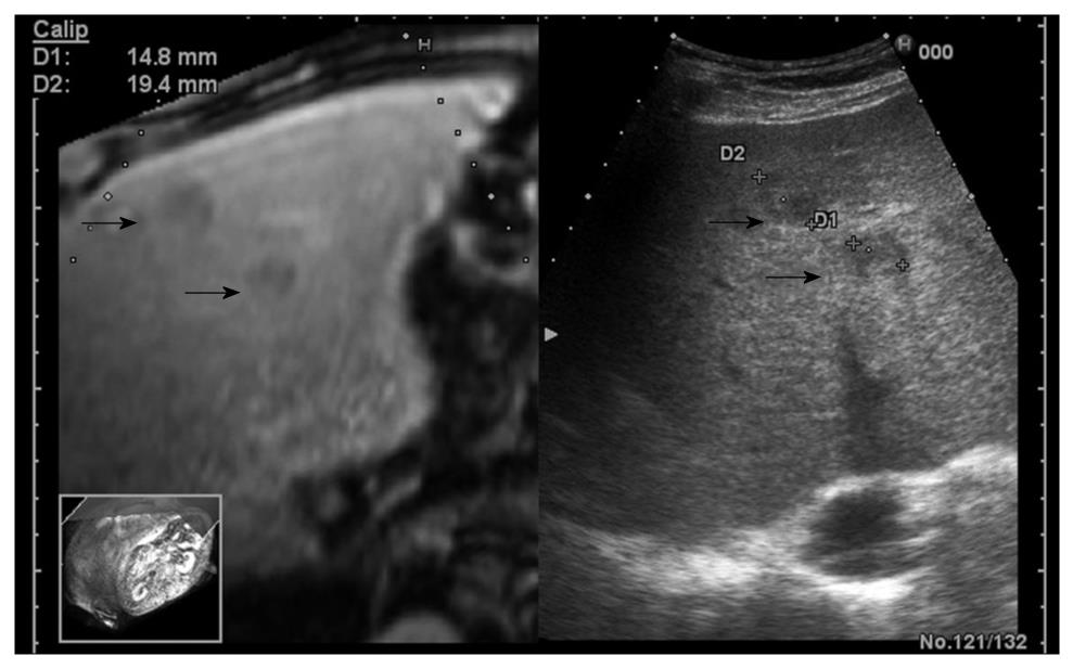
Figure 3 A 65-year-old male patient with liver metastases.
The workstation monitor displays the ultrasound and computed tomography images in real-time.
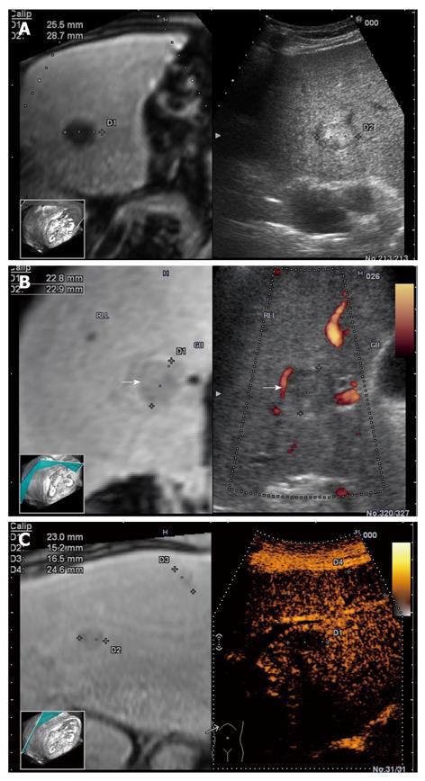
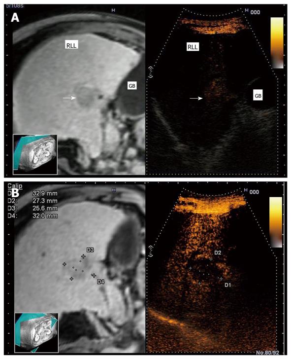
Figure 4 Real-time virtual sonography of liver nodules with simultaneous display of the contrast-enhanced magnetic resonance section and ultrasound images.
2D (A), Power Doppler (B), and contrast enhanced ultrasound (C) vs the reconstructed corresponding image.
Figure 5 Single hepatocellular carcinoma (3 cm) nodule in a cirrhotic patient.
In real-time virtual ultrasonography, after contrast agent injection, a progressive and intense enhancement is observed in the arterial phase (A), followed by wash-out in the tardive phase (B), on contrast-enhanced ultrasound, versus corresponding contrast-enhanced magnetic resonance images.
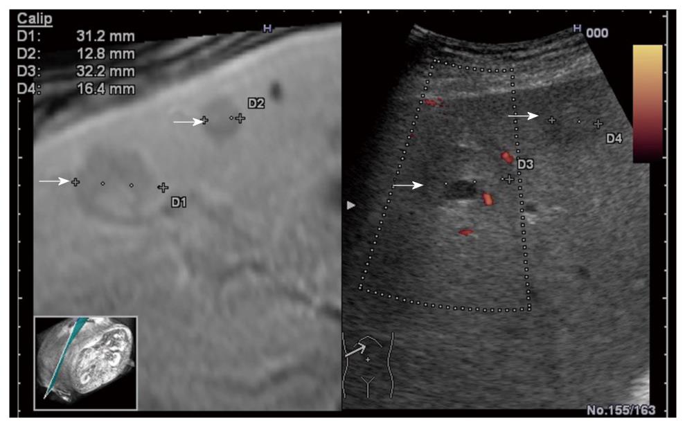
Figure 6 Real-time virtual sonography showing multiple metastases, better depicted on contrast-enhanced computed tomography as compared with ultrasound.
A nodule in nodule aspect is visualized with excellent correlation on both contrast-enhanced magnetic resonance and real-time ultrasound.
- Citation: Sandulescu DL, Dumitrescu D, Rogoveanu I, Saftoiu A. Hybrid ultrasound imaging techniques (fusion imaging). World J Gastroenterol 2011; 17(1): 49-52
- URL: https://www.wjgnet.com/1007-9327/full/v17/i1/49.htm
- DOI: https://dx.doi.org/10.3748/wjg.v17.i1.49


