Copyright
©2011 Baishideng Publishing Group Co.
World J Gastroenterol. Jan 7, 2011; 17(1): 15-20
Published online Jan 7, 2011. doi: 10.3748/wjg.v17.i1.15
Published online Jan 7, 2011. doi: 10.3748/wjg.v17.i1.15
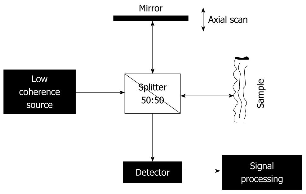
Figure 1 Scheme of a standard optical coherence tomography set-up based on a Michelson interferometer.
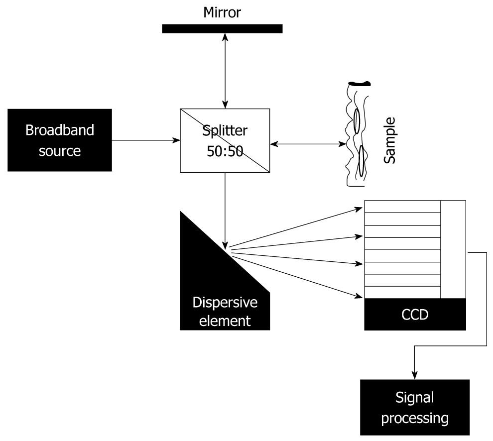
Figure 2 Scheme of fourier domain-optical coherence tomography set-up.
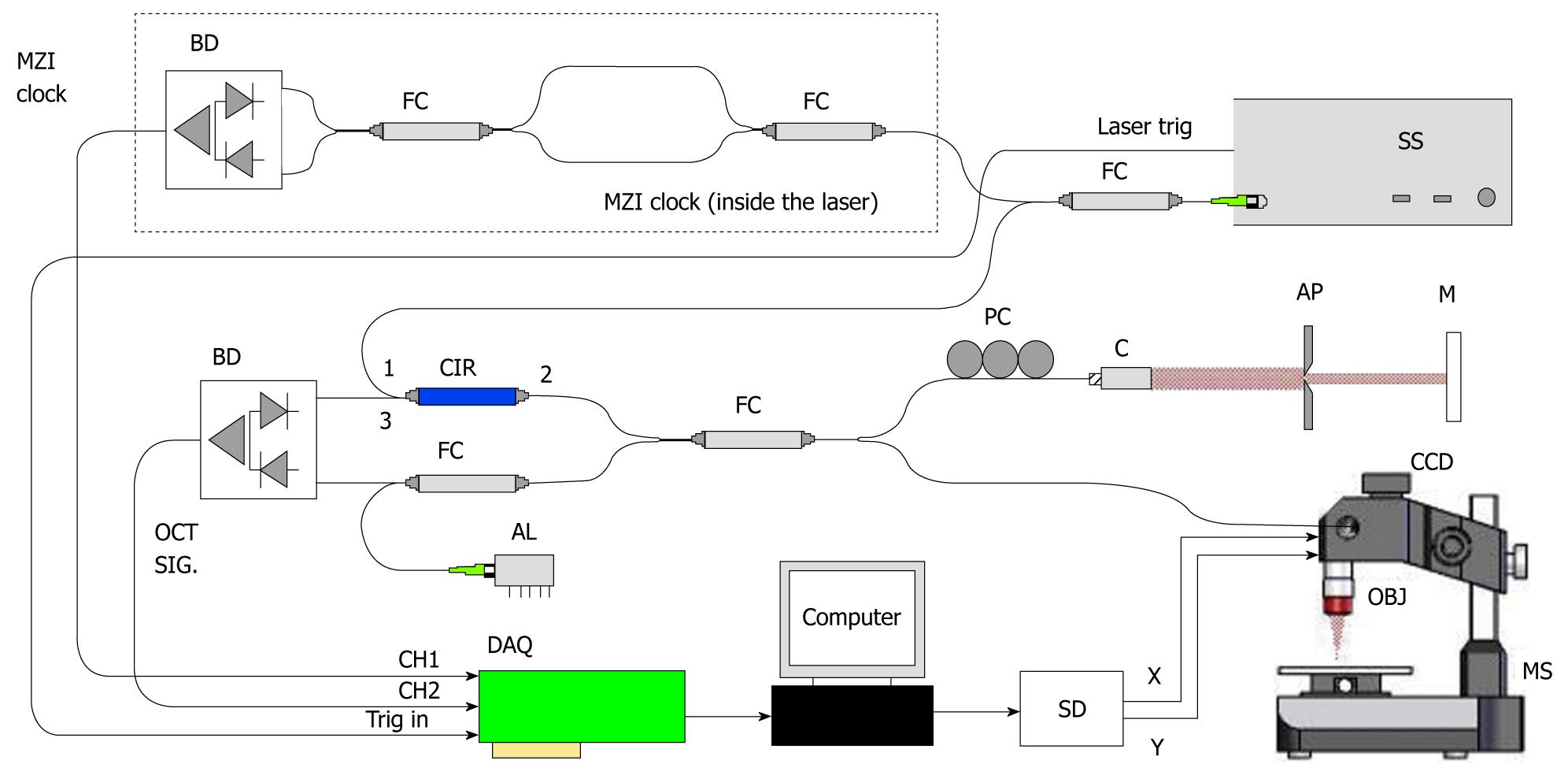
Figure 3 Set-up for OCS1300SS (courtesy of Thorlabs).
OCT: Optical coherence tomography; SS: Swept source.
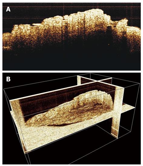
Figure 4 2-D (A) and 3-D (B) optical coherence tomography images of gastric tissue biopsy with visualization of normal components of the parietal layers.
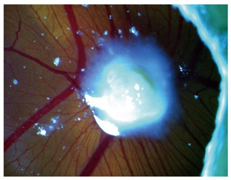
Figure 5 Macroscopic view of a chick embryo chorioallantoic membrane with human gastric tumor implant.
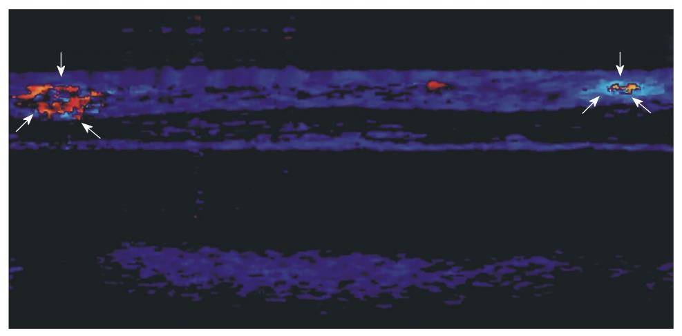
Figure 6 Doppler-optical coherence tomography imaging of a chick embryo chorioallantoic membrane (2 mm × 2 mm) showing capillary activity (arrows) near a human gastric tumor implant.
- Citation: Osiac E, Săftoiu A, Gheonea DI, Mandrila I, Angelescu R. Optical coherence tomography and Doppler optical coherence tomography in the gastrointestinal tract. World J Gastroenterol 2011; 17(1): 15-20
- URL: https://www.wjgnet.com/1007-9327/full/v17/i1/15.htm
- DOI: https://dx.doi.org/10.3748/wjg.v17.i1.15


