Copyright
©2011 Baishideng Publishing Group Co.
World J Gastroenterol. Jan 7, 2011; 17(1): 1-6
Published online Jan 7, 2011. doi: 10.3748/wjg.v17.i1.1
Published online Jan 7, 2011. doi: 10.3748/wjg.v17.i1.1
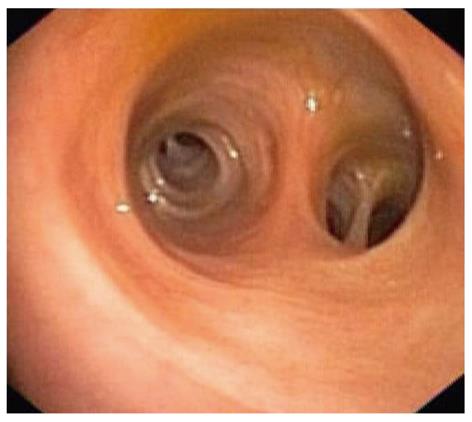
Figure 1 Cholangioscopic view of normal intrahepatic biliary mucosa (image by a prototype video cholangioscope, CHF type Y0002, Olympus Corporation, Tokyo, Japan).
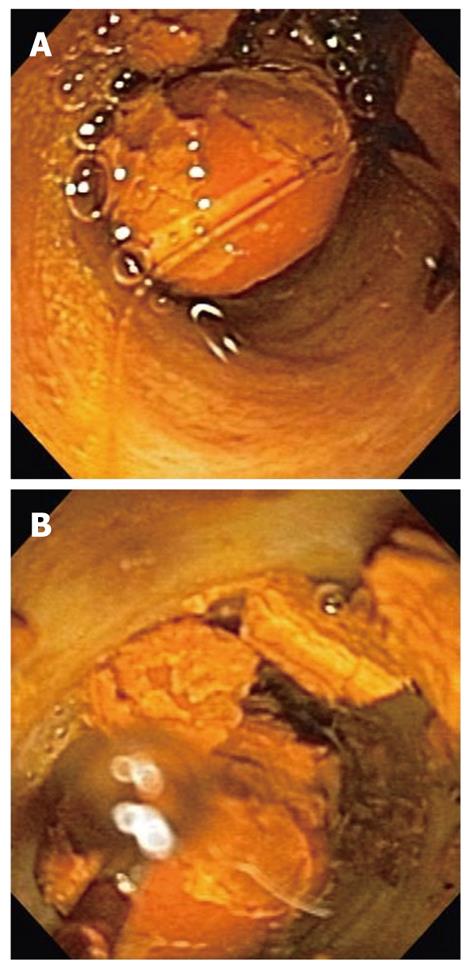
Figure 2 Cholangioscopic views of a bile duct stone prior to (A) and after (B) electrohydraulic lithotripsy.
The lithotripsy probe is visible in the left lower corner of (B).
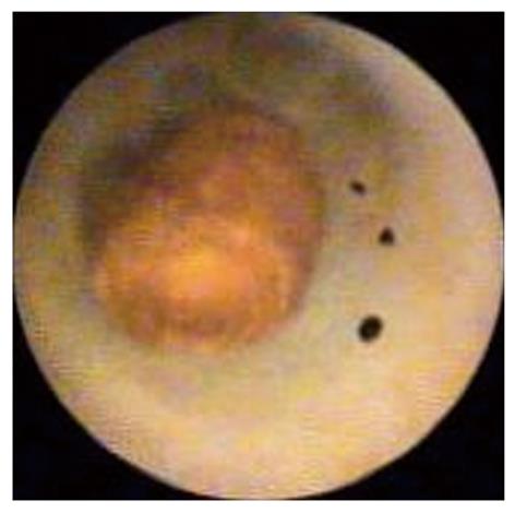
Figure 3 Cholangioscopic view of a small stone surrounded by contrast in an intrahepatic duct.
The stone was missed during endoscopic retrograde cholangiopancreatography (image by Spyglass Direct Visualization System, Boston Scientific, Natick, USA).
- Citation: Parsi MA. Peroral cholangioscopy in the new millennium. World J Gastroenterol 2011; 17(1): 1-6
- URL: https://www.wjgnet.com/1007-9327/full/v17/i1/1.htm
- DOI: https://dx.doi.org/10.3748/wjg.v17.i1.1


