©2010 Baishideng.
World J Gastroenterol. Feb 21, 2010; 16(7): 804-817
Published online Feb 21, 2010. doi: 10.3748/wjg.v16.i7.804
Published online Feb 21, 2010. doi: 10.3748/wjg.v16.i7.804
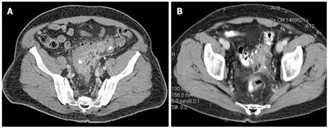
Figure 1 Diverticulitis.
A: Uncomplicated sigmoid diverticulitis with colonic thickening and straining at CT (arrow), also referred to as “mild” CT diverticulitis. Two diverticula contain contrast medium without evidence of extravasation outside the sigmoid; B: “Severe” CT diverticulitis with extravasation of contrast and small amount of extraluminal air (arrow). This patient was initially managed non-operatively and eventually required surgery for recurrent disease.
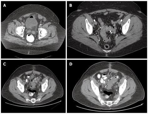
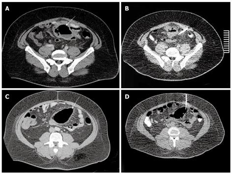
Figure 2 Fistula.
A: Colovesical fistula as indicated by the presence of air in the bladder. This patient had symptoms and other CT findings consistent with sigmoid diverticulitis; B: Sigmoid diverticulitis and colovaginal fistula. This patient had undergone previous hysterectomy and complained of feculent discharge from her vagina. CT scan indicated inflamed sigmoid with adherent small bowel loop (arrow). The small bowel loop could be successfully separated from the sigmoid at the time of laparoscopic sigmoidectomy. There was no evidence of coloenteric fistula; Sigmoid diverticulitis with colocutaneous fistula (arrows) (C and D) (courtesy of Dr. Ravi Pokala Kiran, Department of Colorectal Surgery, Digestive Disease Institute, Cleveland Clinic, Cleveland, Ohio, USA).
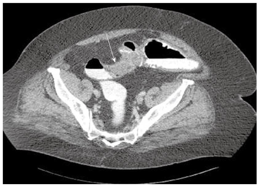
Figure 3 Sigmoid stricture (arrow) causing large bowel obstruction with proximal colonic dilatation.
Clinical and imaging findings at presentation did not allow ruling out sigmoid carcinoma. This patient was treated with initial Hartmann procedure and the pathology report revealed sigmoid diverticulitis. He subsequently underwent Hartmann takedown after 3 mo.
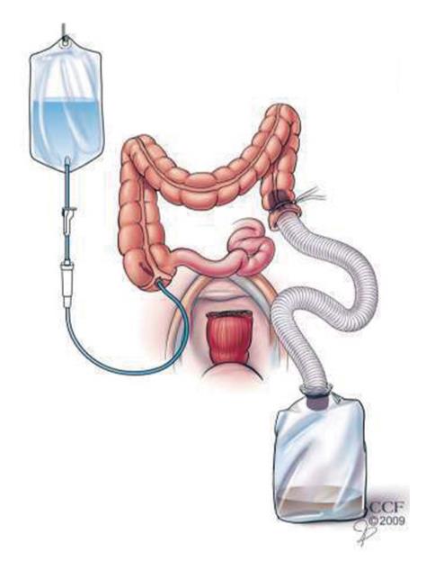
Figure 4 On-table intraoperative colonic lavage (see explanation in text).
Figure 5 Sigmoid diverticulitis complicated by pericolic abscesses (A and C, arrows) requiring treatment by placement of two separate CT-guided percutaneous drains (B and D).
This patient underwent laparoscopic sigmoidectomy with primary colorectal anastomosis and removal of both drains 6 wk after percutaneous drain placement.
- Citation: Stocchi L. Current indications and role of surgery in the management of sigmoid diverticulitis. World J Gastroenterol 2010; 16(7): 804-817
- URL: https://www.wjgnet.com/1007-9327/full/v16/i7/804.htm
- DOI: https://dx.doi.org/10.3748/wjg.v16.i7.804


