Copyright
©2010 Baishideng Publishing Group Co.
World J Gastroenterol. Dec 14, 2010; 16(46): 5822-5829
Published online Dec 14, 2010. doi: 10.3748/wjg.v16.i46.5822
Published online Dec 14, 2010. doi: 10.3748/wjg.v16.i46.5822
Figure 1 Dermoid cyst in a 63-year-old man, who was misdiagnosed as having abscess.
Axial T1-weighted (A), T2-weighted fat-suppression (B) and Sagittal T2-weighted (C) magnetic resonance (MR) images show a thin regular peripheral rim (black arrows) circumscribing a retrorectal cyst and containing some air (white arrows). The cyst lesion extends to the left ischiorectal fossa, and is hypointense on T1-weighted (A) and hyperintense on T2-weighted MR images (B, C).
Figure 2 Tailgut cyst in a 30-year-old man.
On T2-weighted fat-suppression (A, B) and Sagittal T2-weighted images (C), small cysts are composed to form a honeycomb (white arrows) adjacent to a main larger cyst (black arrows).
Figure 3 Tailgut cyst associated with mucinous adenocarcinoma in a 52-year-old woman.
A, B: The rectum is compressed and shifted to the front but without evidence of invasion. The cystic portion (black arrows) is hyperintense, and the malignant portion (white arrows) presents as irregular margin with intermediate signal intensity on T2-weighted fat-suppression (A) and Sagittal T2-weighted images (B); C: Photomicrograph of the tumor shows a small cluster of atypical cells in stroma.
Figure 4 Adenocarcinoma of the anal duct in a 71-year-old woman.
A, B: Axial T1-weighted magnetic resonance (MR) image shows a low signal mass with irregular marginal in the retrorectal space and high signal on T2-weighted MR image; C: The mucin, which forms the major tissue component of mucinous tumor, shows high signal intensity (white arrows) on fat-suppressed T2-weighted image; D: The gland shows irregular features with cellular hyperchromatin and the stroma shows marked desmoplasia.
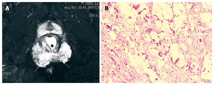
Figure 5 A 59-year-old man with mucinous adenocarcinoma caused by anal fistula.
A: Axial fat-suppressed T2-weighted magnetic resonance image shows a horse-shoe mass with typical mesh-like enhancing areas (arrows). The mass has a internal fistula connected to the anorectum (arrows); B: Microscopy shows a single or small cluster of atypical cells floating in mucin pool and bundles of collagen with hyaline degeneration in stroma.
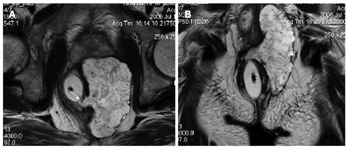
Figure 6 A 52-year-old man with mucinous adenocarcinoma caused by anal fistula.
A, B: The mass displays heterogeneous intensity on T2-weighted magnetic resonance image. There is a fistula between the mass and the anus (black arrow) and an internal opening in anorectum (white arrow).
Figure 7 Primary retrorectal adenocarcinoma in a 33-year-old man.
A, B: The mass displays low signal intensity, without rim (black arrows) on T1-weighted magnetic resonance (MR) image, and high signal intensity of the irregular border (white arrows) on fat-suppressed T2-weighted MR image; C: Microscopy shows irregular glands of variable sizes and atypical tumor cells. The stroma shows desmoplasia.
Figure 8 Gastrointestinal stromal tumors of rectum in a 44-year-old man.
A well-circumscribed, smooth, intramural mass with exophytic growth expanded from the rectal wall and extended to the right ischiorectal fossa. The tumor displays low signal intensity on T1-weighted image (A), and high signal intensity on T2-weighted image (B, C). There are some areas of necrosis (black arrows) and fibrotic tissues (white arrows) in the tumor.
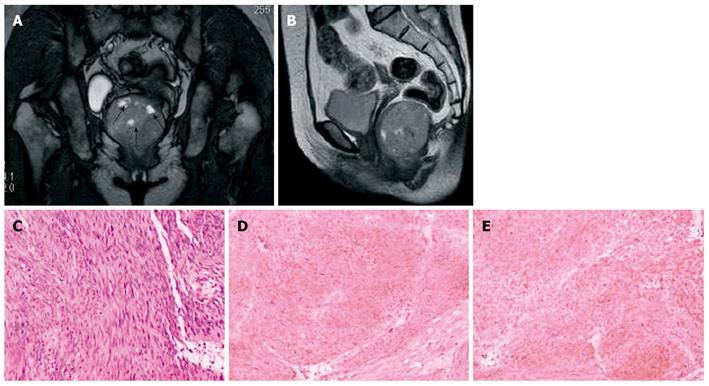
Figure 9 Retrorectal gastrointestinal stromal tumors in a 51-year-old woman.
A, B: T2-weighted magnetic resonance images show a large retrorectal mass, in which some necrosis presents hyperintensity (black arrows); C: Histopathology shows spindle cells and cytoplasmic vacuoles; D, E: On immunohistochemical studies, diffuse and strong immunoreactivity for CD117 and CD34 are seen.
- Citation: Yang BL, Gu YF, Shao WJ, Chen HJ, Sun GD, Jin HY, Zhu X. Retrorectal tumors in adults: Magnetic resonance imaging findings. World J Gastroenterol 2010; 16(46): 5822-5829
- URL: https://www.wjgnet.com/1007-9327/full/v16/i46/5822.htm
- DOI: https://dx.doi.org/10.3748/wjg.v16.i46.5822


