©2010 Baishideng Publishing Group Co.
World J Gastroenterol. Oct 28, 2010; 16(40): 5122-5129
Published online Oct 28, 2010. doi: 10.3748/wjg.v16.i40.5122
Published online Oct 28, 2010. doi: 10.3748/wjg.v16.i40.5122
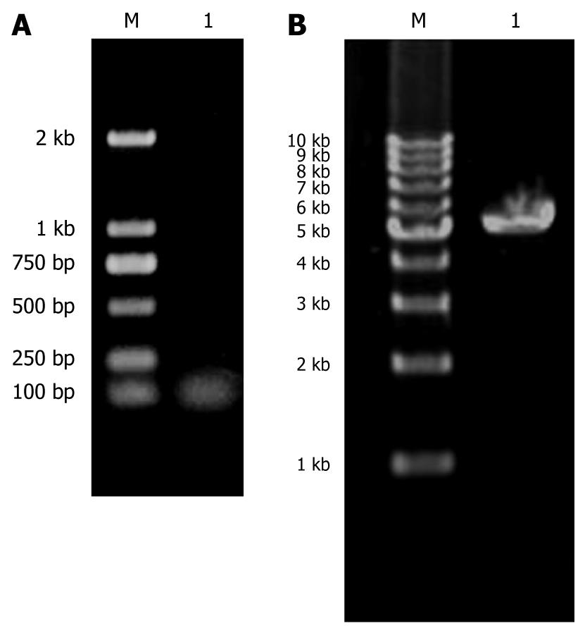
Figure 1 Agarose gel electrophoresis.
A: KIT (B/H) was about 50 bp in length in lane 1. Lane M: DNA Marker DL2000; B: PDC316-EGFP-U6 was about 5300 bp in lane 1, consistent with the vector length. Lane M: 1 kb DNA Ladder Marker.
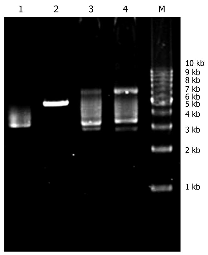
Figure 2 After recombination, restrictive endonuclease site SalI was eliminated.
The blank plasmid could be linearized by SalI digestion, while the recombinant one could not. The plasmids on lanes 3 and 4 were the recombinant PDC316-EGFP-U6-KIT. Lane 1: Blank PDC316-EGFP-U6; Lane 2: Blank PDC316-EGFP-U6 digested with SalI; Lane 3: Recombinant PDC316-EGFP-U6-KIT; Lane 4: Recombinant PDC316-EGFP-U6-KIT digested with SalI; Lane M: DNA Marker DL2000.
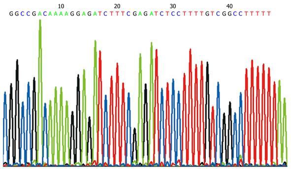
Figure 3 Sequencing graph shows that the KIT RNAi sequence in PDC316-EGFP-U6-KIT plasmid was correct.
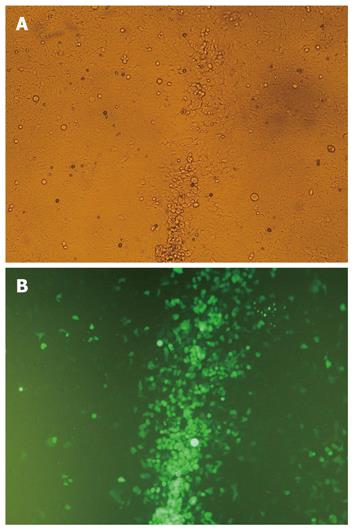
Figure 4 Cytopathic effect and green fluorescence in HEK293 cells observed after recombination of PDC316-EGFP-U6-KIT and pBHGlox(delta) E1,3Cre.
A: Cytopathic effect of HEK293; B: Fluorescence in HEK293 cell, × 200.
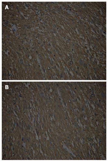
Figure 5 Positive staining of CD117 in gastrointestinal stromal tumor.
A: Primary gastric gastrointestinal stromal tumor; B: Xenograft, × 200.
- Citation: Wang TB, Huang WS, Lin WH, Shi HP, Dong WG. Inhibition of KIT RNAi mediated with adenovirus in gastrointestinal stromal tumor xenograft. World J Gastroenterol 2010; 16(40): 5122-5129
- URL: https://www.wjgnet.com/1007-9327/full/v16/i40/5122.htm
- DOI: https://dx.doi.org/10.3748/wjg.v16.i40.5122


