©2010 Baishideng.
World J Gastroenterol. Jan 28, 2010; 16(4): 496-500
Published online Jan 28, 2010. doi: 10.3748/wjg.v16.i4.496
Published online Jan 28, 2010. doi: 10.3748/wjg.v16.i4.496
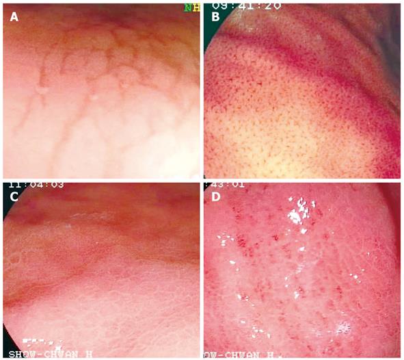
Figure 1 Mucosal patterns.
A: Type 1 mucosal pattern showing cleft-like appearance; B: Type 2 mucosal pattern showing regular arrangement of red dots; C: Type 3 mucosal pattern showing mosaic appearance; D: Type 4 mucosal pattern showing mosaic appearance with focal area of hyperemia.
-
Citation: Yan SL, Wu ST, Chen CH, Hung YH, Yang TH, Pang VS, Yeh YH. Mucosal patterns of
Helicobacter pylori -related gastritis without atrophy in the gastric corpus using standard endoscopy. World J Gastroenterol 2010; 16(4): 496-500 - URL: https://www.wjgnet.com/1007-9327/full/v16/i4/496.htm
- DOI: https://dx.doi.org/10.3748/wjg.v16.i4.496


