©2010 Baishideng.
World J Gastroenterol. Jan 28, 2010; 16(4): 484-489
Published online Jan 28, 2010. doi: 10.3748/wjg.v16.i4.484
Published online Jan 28, 2010. doi: 10.3748/wjg.v16.i4.484
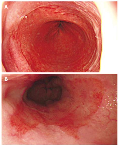
Figure 1 Shape of Barrett’s epithelium.
We originally divided Barrett’s epithelium into two types based on its shape. A: L type, in which the difference between the C extent and M extent was < 2 cm and the visible red columnar epithelium could be observed as a lotus-like shape; B: F type, in which the difference was ≥ 2 cm and the columnar epithelium of Barrett’s epithelium was observed as a flame-like shape.
- Citation: Akiyama T, Inamori M, Iida H, Endo H, Hosono K, Sakamoto Y, Fujita K, Yoneda M, Takahashi H, Koide T, Tokoro C, Goto A, Abe Y, Shimamura T, Kobayashi N, Kubota K, Saito S, Nakajima A. Shape of Barrett’s epithelium is associated with prevalence of erosive esophagitis. World J Gastroenterol 2010; 16(4): 484-489
- URL: https://www.wjgnet.com/1007-9327/full/v16/i4/484.htm
- DOI: https://dx.doi.org/10.3748/wjg.v16.i4.484


