Copyright
©2010 Baishideng Publishing Group Co.
World J Gastroenterol. Sep 14, 2010; 16(34): 4253-4263
Published online Sep 14, 2010. doi: 10.3748/wjg.v16.i34.4253
Published online Sep 14, 2010. doi: 10.3748/wjg.v16.i34.4253
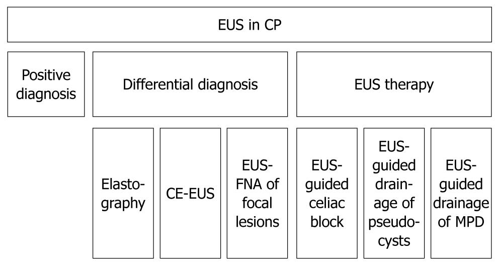
Figure 1 Flowchart of the endoscopic ultrasonography utility in chronic pancreatitis.
EUS: Endoscopic ultrasonography; CP: Chronic pancreatitis; CE-EUS: Contrast enhanced endoscopic ultrasonography; MPD: Main pancreatic duct.
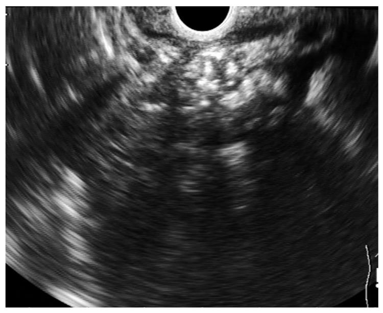
Figure 2 Chronic pancreatitis.
Parenchymal and ductal pancreatic stones as hyperechoic structures with shadowing and stenosis of the main pancreatic duct.
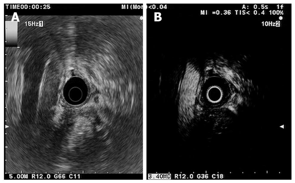
Figure 3 Mass resembling chronic pancreatitis.
A: Conventional endoscopic ultrasonography (EUS). Hypoechoic inhomogeneous mass in the pancreatic head. Aorta and inferior caval vein are also seen; B: Contrast-enhanced harmonic-EUS. During the arterial phase (25 s after contrast injection) the abdominal aorta becomes hyperechoic and the mass is hypovascular compared with surrounding parenchyma.
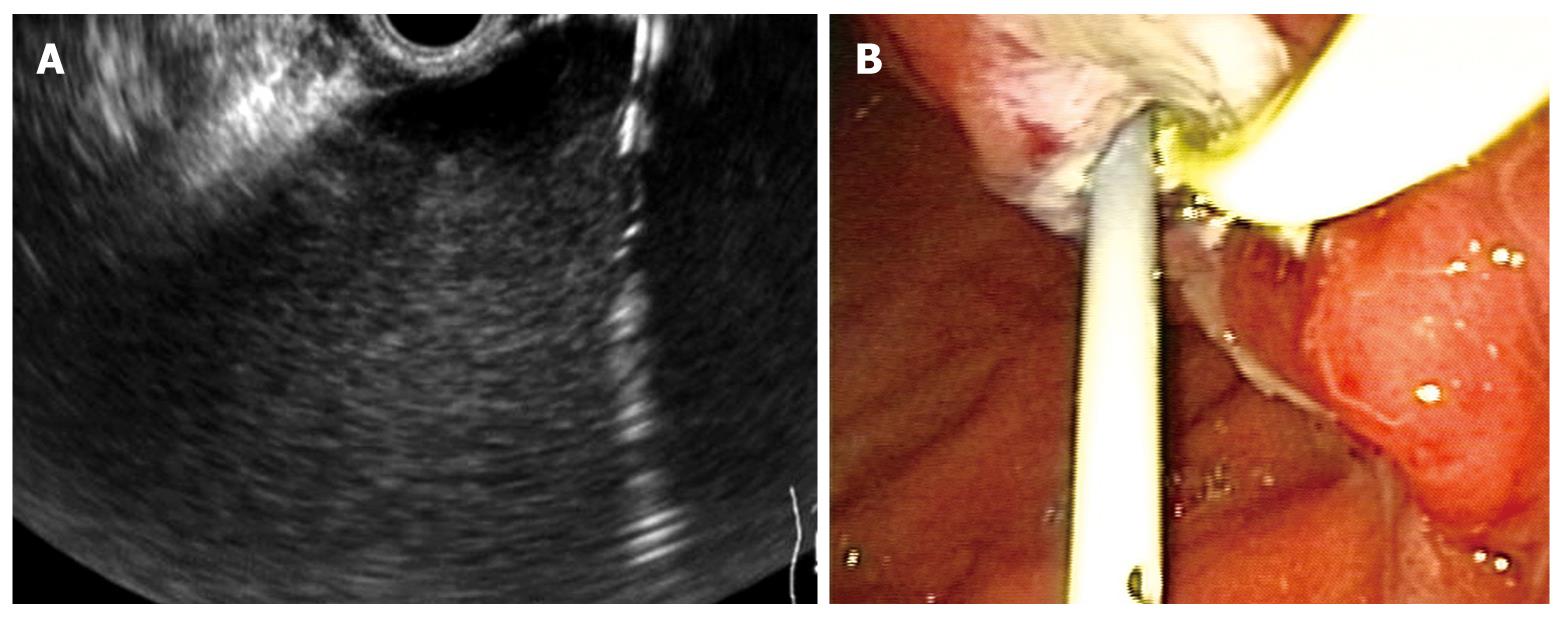
Figure 4 Endoscopic ultrasonography-guided pseudocyst drainage.
A: The cystostomy is seen as a hyperechoic parallel structure inside the hypoechoic well-delineated pseudocyst; B: Endoscopic view of a stent and a nasocystic drainage placed transgastric into a pseudocyst.
- Citation: Seicean A. Endoscopic ultrasound in chronic pancreatitis: Where are we now? World J Gastroenterol 2010; 16(34): 4253-4263
- URL: https://www.wjgnet.com/1007-9327/full/v16/i34/4253.htm
- DOI: https://dx.doi.org/10.3748/wjg.v16.i34.4253


