©2010 Baishideng.
World J Gastroenterol. Aug 21, 2010; 16(31): 3984-3986
Published online Aug 21, 2010. doi: 10.3748/wjg.v16.i31.3984
Published online Aug 21, 2010. doi: 10.3748/wjg.v16.i31.3984
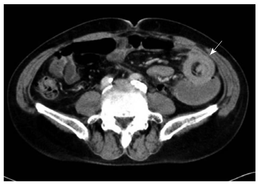
Figure 1 Abdominal computed tomography discloses a typical “target sign” over the small intestine (arrow).
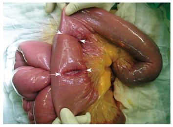
Figure 2 Intraoperative photo showing intussusceptions in the small intestine (arrows).
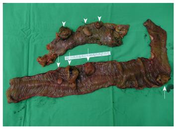
Figure 3 Surgical specimen reveals metastatic localized malignant pleural mesothelioma in the small intestine (arrows) and colon (arrow heads).
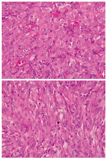
Figure 4 Microscopic examination reveals the tumor to consist of sheets of spindle to epithelioid cells with abundant eosinophilic cytoplasm and vesicular nuclear chromatin with prominent nucleoli (HE stain, × 400).
- Citation: Liu H, Cheng YJ, Chen HP, Hwang JC, Chang PC. Multiple bowel intussusceptions from metastatic localized malignant pleural mesothelioma: A case report. World J Gastroenterol 2010; 16(31): 3984-3986
- URL: https://www.wjgnet.com/1007-9327/full/v16/i31/3984.htm
- DOI: https://dx.doi.org/10.3748/wjg.v16.i31.3984


