©2010 Baishideng.
World J Gastroenterol. Aug 21, 2010; 16(31): 3928-3935
Published online Aug 21, 2010. doi: 10.3748/wjg.v16.i31.3928
Published online Aug 21, 2010. doi: 10.3748/wjg.v16.i31.3928
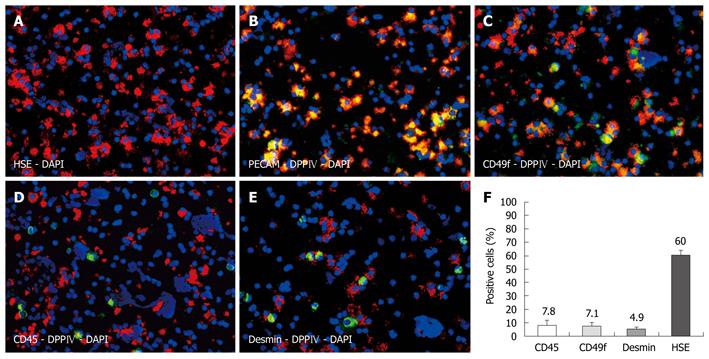
Figure 1 Characterization of non-parenchymal cell preparations by immunofluorescence on cytospins.
Hepatic sinusoidal endothelial (HSE) (red) identifying approximately 60% of all cells as hepatic sinusoidal cells (A), donor specific dipeptidyl peptidase IV (DPPIV) (red) co-localized with platelet endothelial cell adhesion molecule (green) (B), and with CD49f as bile duct marker (green) (C). Donor cells were negative for CD45 (D) and desmin (E). Labeling indices (F), nuclear counterstaining with DAPI (blue), original magnification × 200.
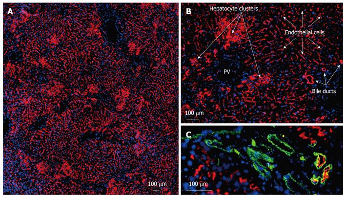
Figure 2 Characteristics of dipeptidyl peptidase IV-positive cells in the liver following transplantation of non-parenchymal cell preparations and hepatocytes.
Frozen sections were obtained from recipient livers present at 16 wk post-transplantation. Acetone-fixed frozen sections were single or double-labeled by indirect immunofluorescence analysis with an antibody detecting the donor cell specific dipeptidyl peptidase IV (red) (A-C) and bile-duct specific anti-CD49f (green) (C). Merged images are combined with blue nuclear DAPI-staining. Circumscribed and compact clusters of hepatocyte repopulation could be distinguished from large areas of endothelial repopulation which covered a maximum of 90% of the section plane. Formation of bile duct-like structures could be detected by co-staining with the duct marker CD49f. PV: Portal vein.
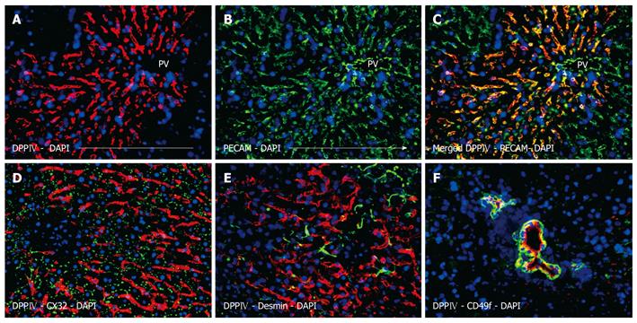
Figure 3 Phenotypic assessment of the non-hepatocytes liver repopulation showing extensive and string-like repopulation by liver sinusoidal endothelial cells emerging from the portal veins.
Donor derived cells were identified by dipeptidyl peptidase IV (DPPIV) immunofluorescence staining (red) (A), which co-localized with the endothelial marker PECAM (green) (B), overlay (C). Donor endothelial cells could be clearly distinguished from endogenous hepatocytes which were outlined by the hepatic differentiation marker CX32 (green) (D), desmin-positive cells (green) were detectable with a regular staining pattern for normal (unharmed) liver (E). Additionally, DPPIV-positive donor cells formed bile duct structures which expressed the specific marker CD49f (green) (C), nuclear counterstaining with DAPI (blue), original magnification × 200.
- Citation: Krause P, Rave-Fränk M, Wolff HA, Becker H, Christiansen H, Koenig S. Liver sinusoidal endothelial and biliary cell repopulation following irradiation and partial hepatectomy. World J Gastroenterol 2010; 16(31): 3928-3935
- URL: https://www.wjgnet.com/1007-9327/full/v16/i31/3928.htm
- DOI: https://dx.doi.org/10.3748/wjg.v16.i31.3928


