Copyright
©2010 Baishideng.
World J Gastroenterol. Jul 14, 2010; 16(26): 3343-3346
Published online Jul 14, 2010. doi: 10.3748/wjg.v16.i26.3343
Published online Jul 14, 2010. doi: 10.3748/wjg.v16.i26.3343
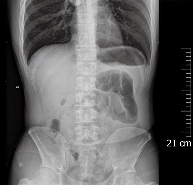
Figure 1 Plain abdominal X-ray.
Mildly dilated pneumatic intestinal canal loops but no typical air-fluid level shadow in the left upper quadrant of abdomen.
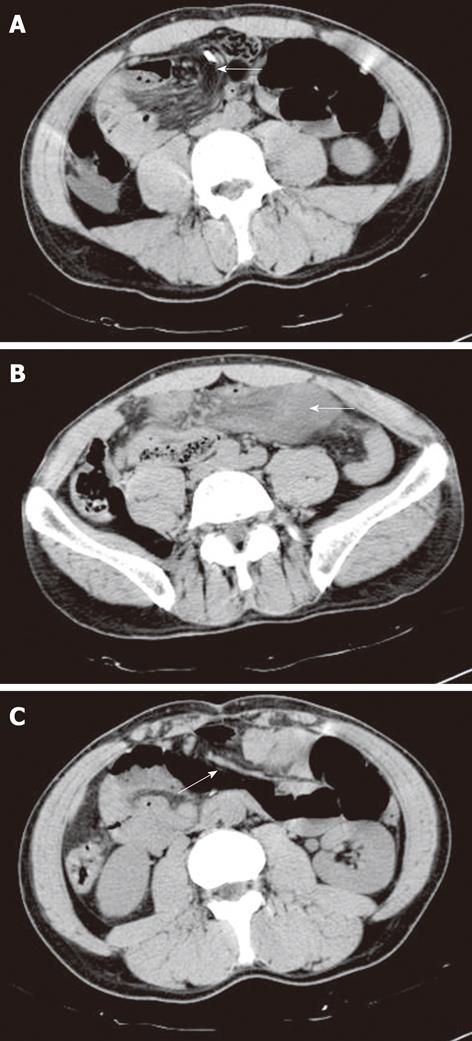
Figure 2 Computed tomography scan.
Suspicious upper abdominal mesentery intorsion (A, arrow), right lower quadrant intestinal loop (B, arrow), middle abdominal line separation (C, arrow).
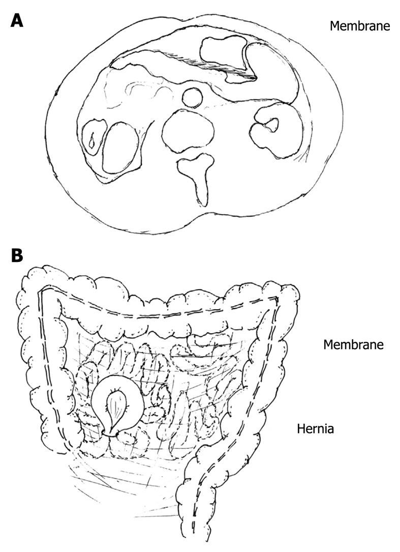
Figure 3 Schema showing a membrane separating the peritoneal cavity into two compartments (A) and descending mesocolon dorsal root connecting with the ascending colon (B).
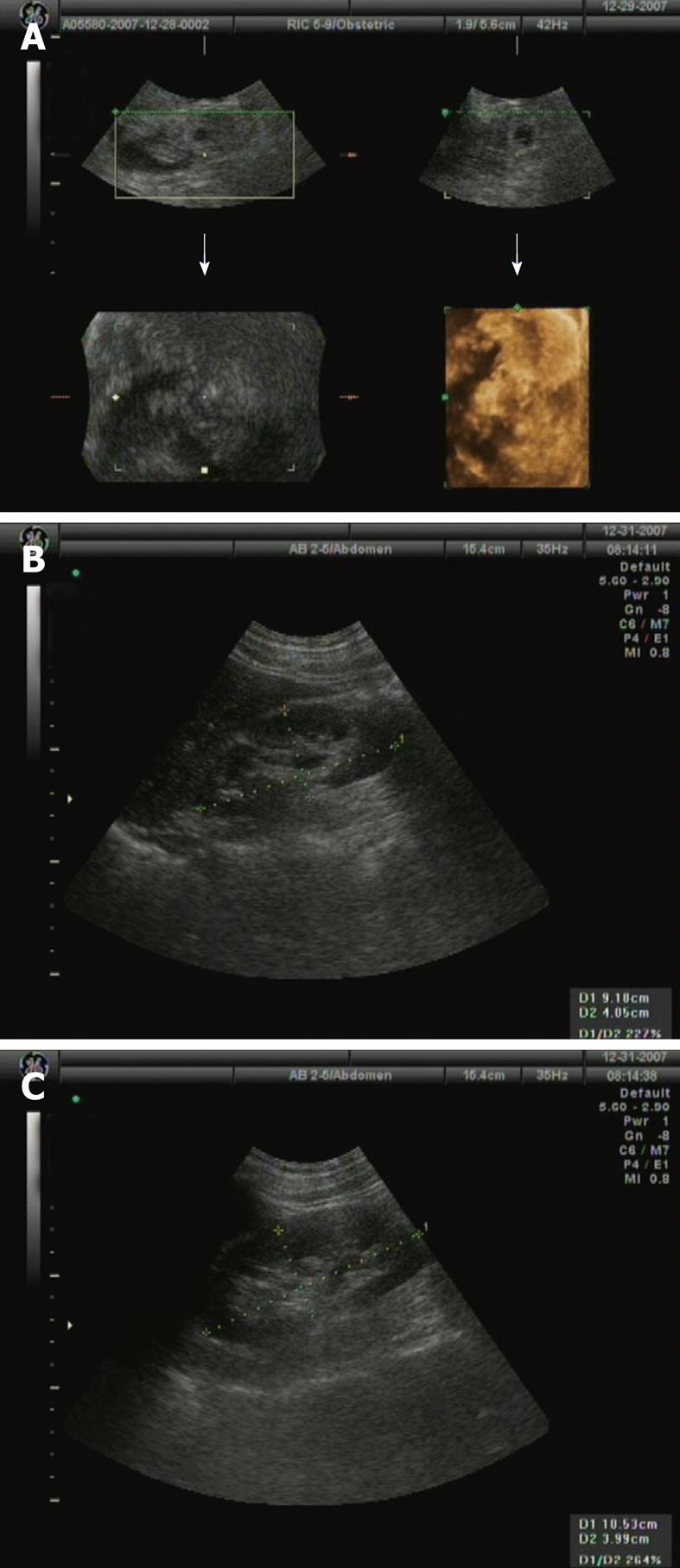
Figure 4 Abdominal and kidney ultrasonography showing no mass and fluid collection (A), left (B) and right (C) normal kidney structure.
- Citation: Liu BL, Chen Y, Liu SQ, Zhang XB, Cui DX, Dai XW. Abdominal separation in an adult male patient with acute abdominal pain. World J Gastroenterol 2010; 16(26): 3343-3346
- URL: https://www.wjgnet.com/1007-9327/full/v16/i26/3343.htm
- DOI: https://dx.doi.org/10.3748/wjg.v16.i26.3343


