Copyright
©2010 Baishideng.
World J Gastroenterol. Jul 14, 2010; 16(26): 3335-3338
Published online Jul 14, 2010. doi: 10.3748/wjg.v16.i26.3335
Published online Jul 14, 2010. doi: 10.3748/wjg.v16.i26.3335
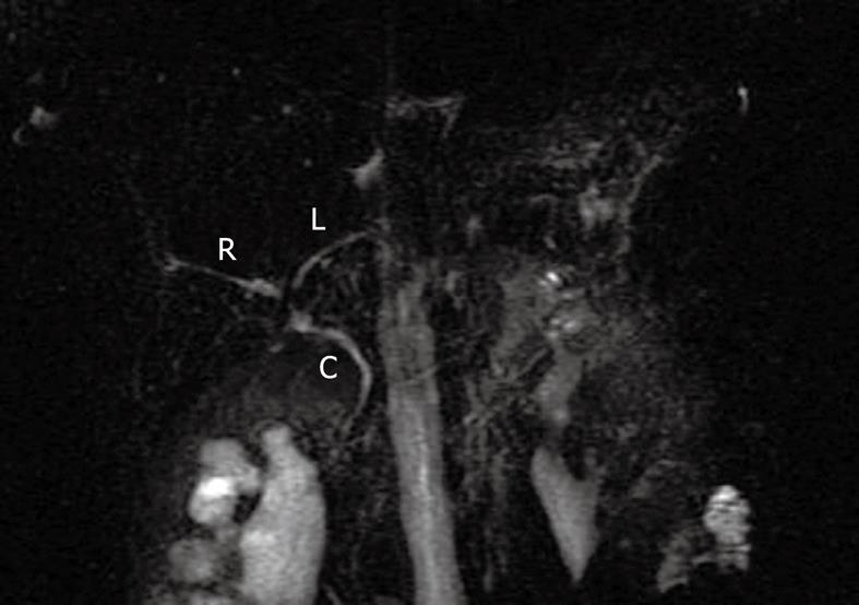
Figure 1 Magnetic resonance cholangiopancreatography.
Patent biliary system without evidence of strictures or obstruction with normal caliber common bile duct (C), right hepatic duct (R), and left hepatic duct (L).
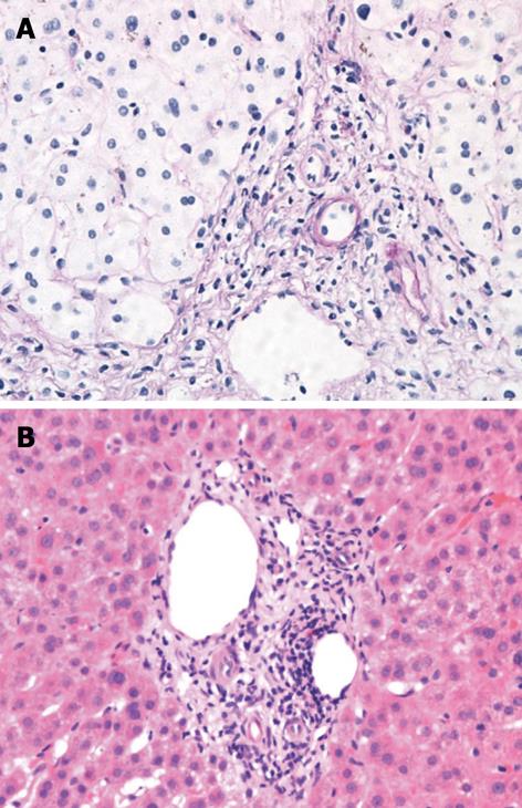
Figure 2 Liver biopsy showing portal tract with normal appearing hepatic vein and hepatic artery, and absence of bile ducts.
A: PAS-D stain, × 200 magnification; B: Hematoxylin and eosin stain, × 200 magnification.
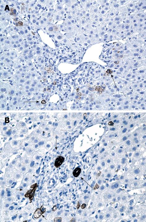
Figure 3 Liver biopsy specimen with cytokeratin staining for biliary elements.
A: No bile ducts are seen, minimal ductular proliferation is apparent at the edges of the portal tract (long arrows); B: Solitary portal tract with presence of irregular bile ducts (long arrows) and ductular proliferation (short arrows).
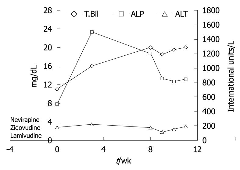
Figure 4 Trend of liver enzymes and bilirubin during the patient’s clinical course.
- Citation: Kochar R, Nevah MI, Lukens FJ, Fallon MB, Machicao VI. Vanishing bile duct syndrome in human immunodeficiency virus: Nevirapine hepatotoxicity revisited. World J Gastroenterol 2010; 16(26): 3335-3338
- URL: https://www.wjgnet.com/1007-9327/full/v16/i26/3335.htm
- DOI: https://dx.doi.org/10.3748/wjg.v16.i26.3335


