Copyright
©2010 Baishideng.
World J Gastroenterol. Jul 14, 2010; 16(26): 3249-3257
Published online Jul 14, 2010. doi: 10.3748/wjg.v16.i26.3249
Published online Jul 14, 2010. doi: 10.3748/wjg.v16.i26.3249
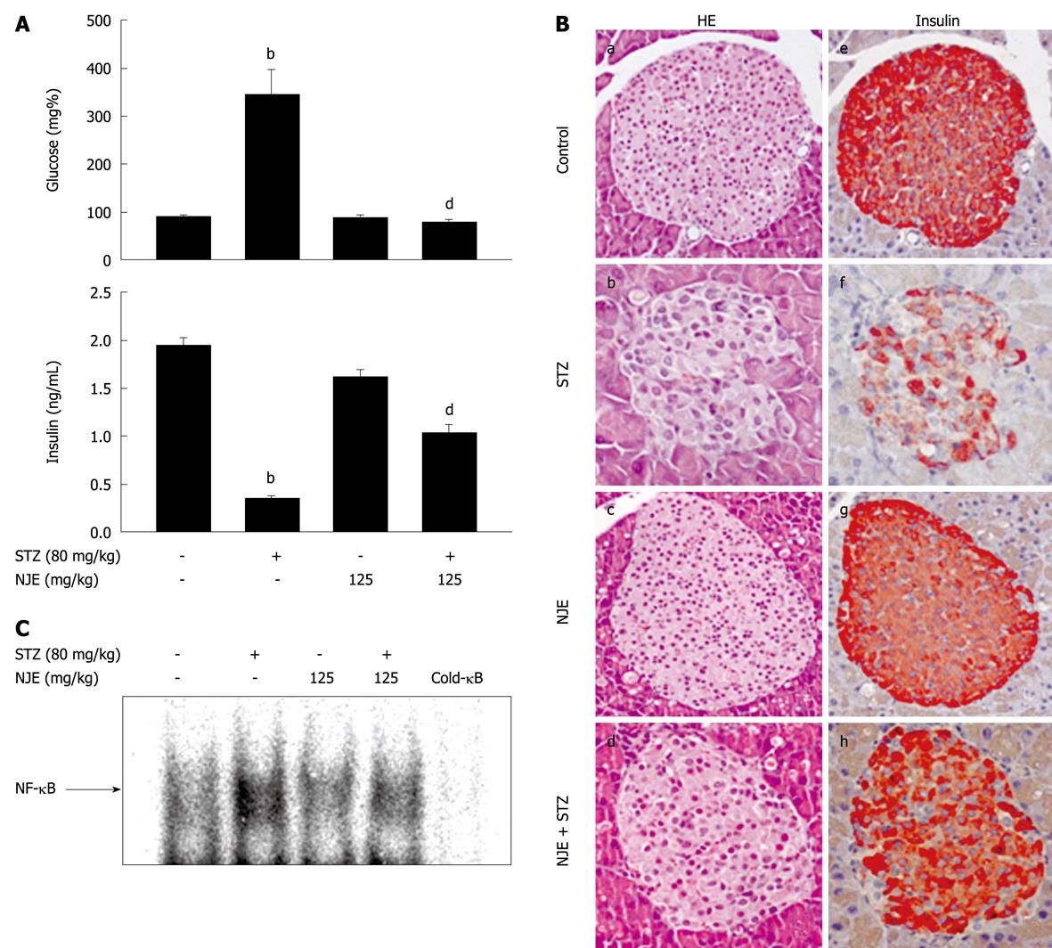
Figure 1 NJE protects islets from STZ-induced destruction.
A: ICR mice were intraperitoneally injected daily with NJE at 125 mg/kg for 3 d and then injected with STZ (80 mg/kg) intravenously. Levels of fasting glucose and insulin were determined; B: Pancreases were obtained from normal controls (a, e), and STZ-injected (b, f), NJE-injected (c, g), and NJE and STZ-injected mice (d, h). Islets and adjoining exocrine regions were counterstained with HE (a-d). Islets were labeled with insulin antibody and peroxidase-labeled anti-rabbit IgG (e-h); C: Nuclear extracts from pancreatic tissues were prepared 30 min after STZ injection, and NF-κB DNA binding was analyzed using electrophoretic mobility shift assay. bP < 0.01 vs untreated control; dP < 0.01 vs STZ-injected group. NJE: Nardostachys jatamansi extract; STZ: Streptozotocin; NF-κB: Nuclear factor κB.
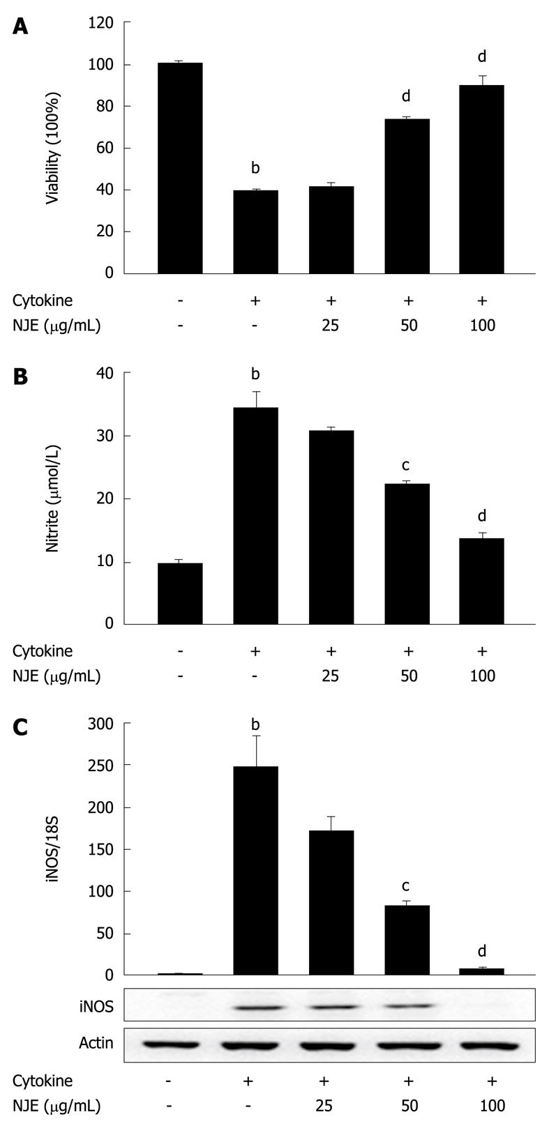
Figure 2 NJE prevents cytokine-induced cell death in RINm5F cells.
A: RINm5F cells were pretreated with NJE for 3 h, and IL-1β (1 U/mL) and IFN-γ (100 U/mL) were added for 48 h. Cell viability was determined using an 3-(4,5-dimethylthiazol-2-yl)-2,5-diphenyltetrazolium bromide assay; B and C: RINm5F cells were pretreated with NJE for 3 h, and IL-1β and IFN-γ were added. Following 24 h of incubation, the level of nitrite production and iNOS mRNA and protein expression were determined. Each value is the mean ± SE of three independent experiments. bP < 0.01 vs untreated controls; cP < 0.05, dP < 0.01 vs cytokine. IL-1β: Interleukin 1β; IFN-γ: Interferon γ.
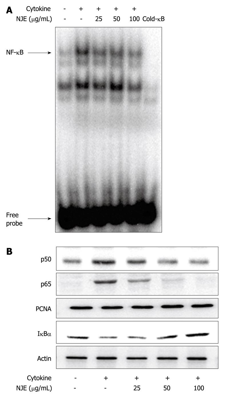
Figure 3 NJE inhibits cytokine-induced NF-κB activation in RINm5F cells.
RINm5F cells were pretreated with NJE for 3 h, and IL-1β (1 U/mL) and IFN-γ (100 U/mL) were added. After 30 min, NF-κB DNA binding was analyzed by EMSA (A), and translocation of p65 and p50 to the nucleus and IκBα degradation in the cytosol (B) were determined by Western blotting. β-actin and PCNA were used as loading controls for cytosolic and nuclear proteins, respectively.
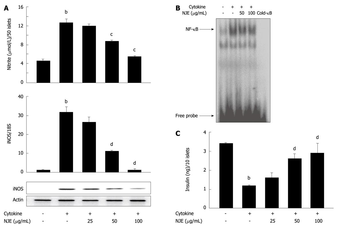
Figure 4 NJE inhibits cytokine-induced activation of NF-κB and maintains glucose-stimulated insulin secretion in rat islets.
Rat islets were treated with IL-1β (1 U/mL) and IFN-γ (100 U/mL) with or without 3 h pretreatment with NJE. Nitrite production and iNOS mRNA and protein expression (A) were determined after 24 h, and NF-κB DNA binding (B) was determined 1 h later; C: Rat islets (10 islets/500 μL) were treated with IL-1β (1 U/mL) and IFN-γ (100 U/mL) with or without 3 h pretreatment with NJE. Following 24 h incubation, glucose-stimulated insulin secretion was quantified. The results of triplicate samples are expressed as the mean ± SE. bP < 0.01 vs untreated controls; cP < 0.05, dP < 0.01 vs cytokine.
-
Citation: Song MY, Bae UJ, Lee BH, Kwon KB, Seo EA, Park SJ, Kim MS, Song HJ, Kwon KS, Park JW, Ryu DG, Park BH.
Nardostachys jatamansi extract protects against cytokine-induced β-cell damage and streptozotocin-induced diabetes. World J Gastroenterol 2010; 16(26): 3249-3257 - URL: https://www.wjgnet.com/1007-9327/full/v16/i26/3249.htm
- DOI: https://dx.doi.org/10.3748/wjg.v16.i26.3249


