Copyright
©2010 Baishideng.
World J Gastroenterol. Jun 28, 2010; 16(24): 3040-3048
Published online Jun 28, 2010. doi: 10.3748/wjg.v16.i24.3040
Published online Jun 28, 2010. doi: 10.3748/wjg.v16.i24.3040
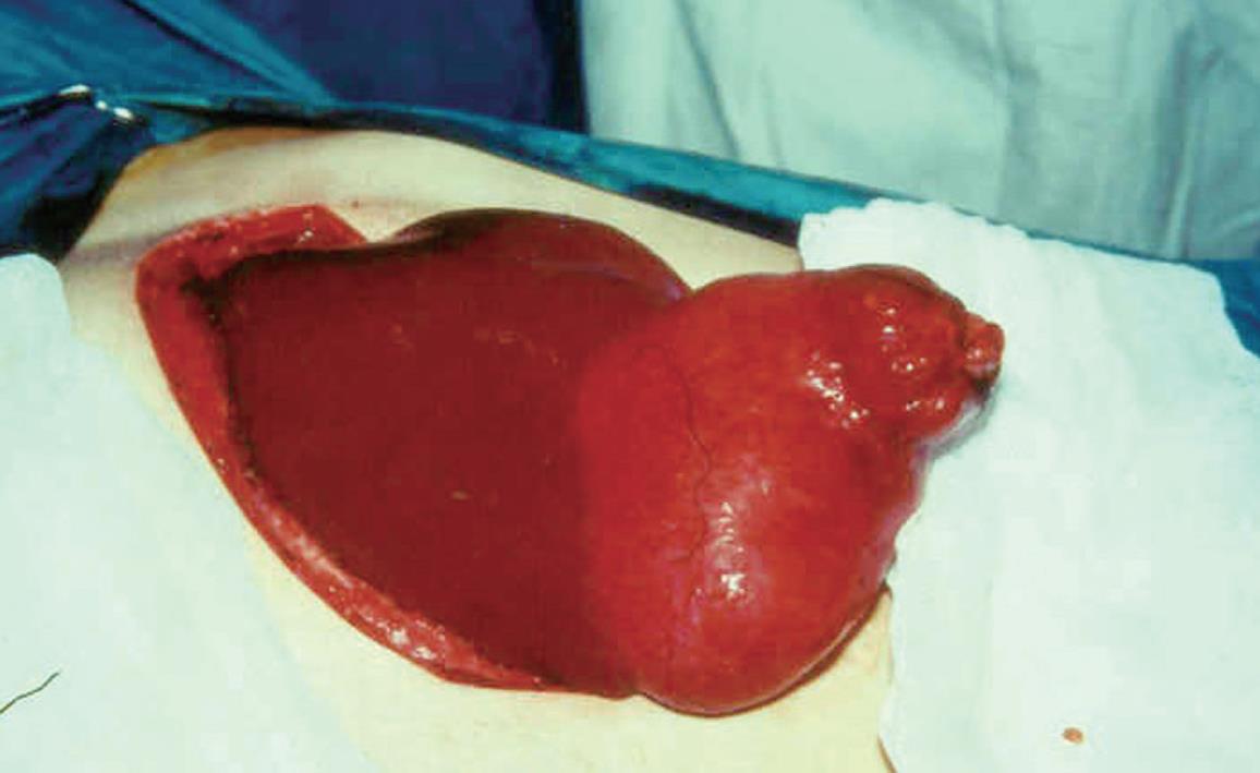
Figure 1 Macroscopic appearance of an hydatid cyst that originates in the right lobe of the liver.
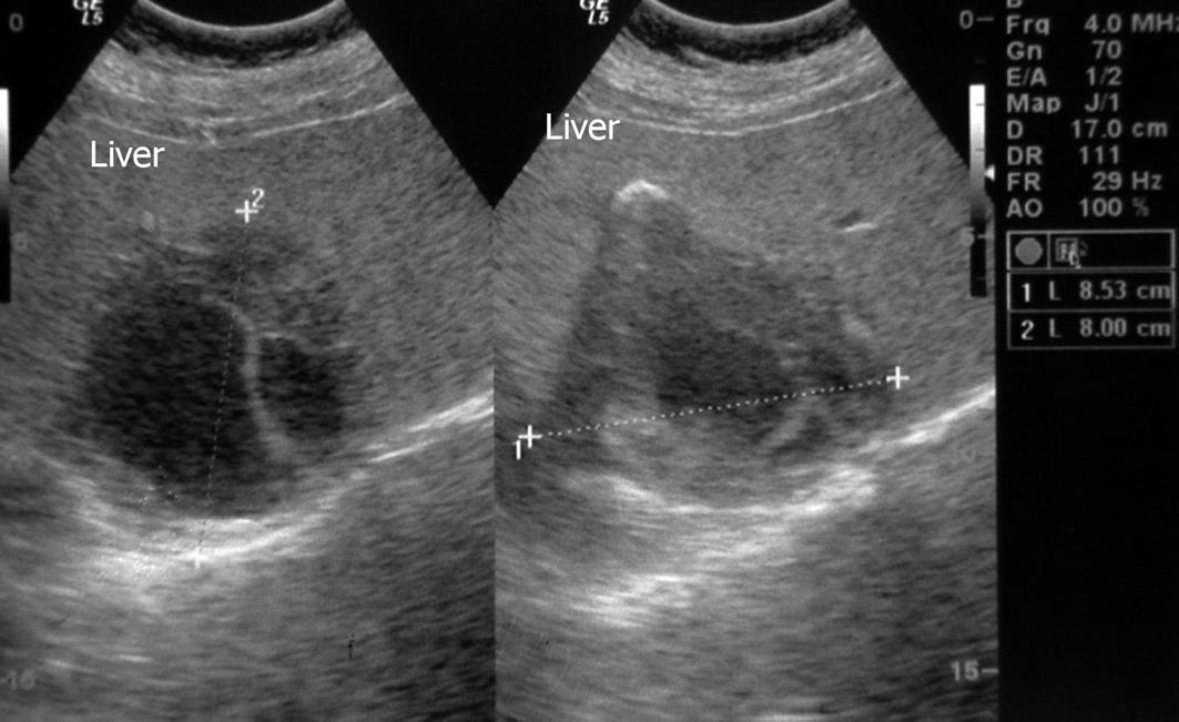
Figure 2 Ultrasonographic appearance of an hydatid cyst of the liver.
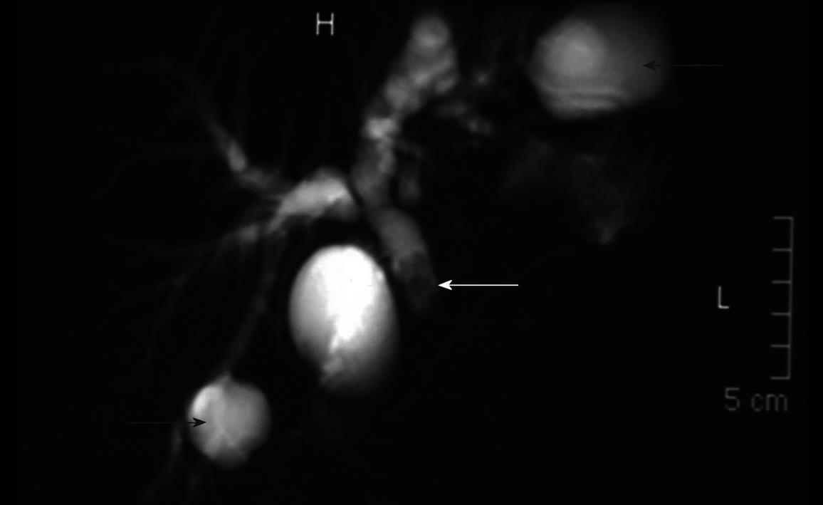
Figure 3 Magnetic resonance imaging cholangiography.
Two biliary ruptured (right and left lobe) hydatid cysts (black arrows) and daughter vesicles in common bile duct (white arrow).
- Citation: Akcan A, Sozuer E, Akyildiz H, Ozturk A, Atalay A, Yilmaz Z. Predisposing factors and surgical outcome of complicated liver hydatid cysts. World J Gastroenterol 2010; 16(24): 3040-3048
- URL: https://www.wjgnet.com/1007-9327/full/v16/i24/3040.htm
- DOI: https://dx.doi.org/10.3748/wjg.v16.i24.3040


